Phenotypic Skin Disease Models
Our advanced phenotypic skin assays leverage healthy human skin biopsies sourced from cosmetic surgery procedures to simulate disease-like phenotypes. By applying proprietary cocktails, we stimulate specific disease pathways in these tissue samples, inducing the upregulation of disease-relevant inflammatory mediators such as Th17 related cytokines in our model of psoriasis. This allows us to model a wide range of skin diseases, including psoriasis, atopic dermatitis, and acne, in a controlled laboratory environment.
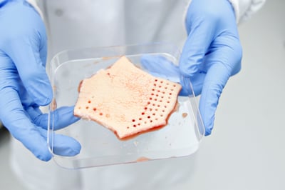
Image: Fresh skin tissue is used to create healthy punch biopsies for our phenotypic disease models.
Key Features of Our Phenotypic Skin Assays
- Disease Simulation in Healthy Skin: Our dedicated R&D team can recreate common disease phenotypes by exposing healthy skin tissue to disease-causing agents. This method activates specific pathways relevant to various skin disorders, enabling the study of disease mechanisms and the testing of potential treatments.
- T-Cell Driven Inflammatory Disease Model: For exploring T-cell driven inflammatory diseases, we utilize a Phytohemagglutinin (PHA) stimulated model. This model activates T-cells and results in the increased release of cytokines such as IL-6, IL-10, IL-13, TNFɑ, and GM-CSF, providing a robust platform for testing anti-inflammatory compounds.
- Inflammatory Acne Phenotype Model: For disease interests specific to inflammatory acne, our Lipopolysaccharide (LPA) stimulated model mimics acne-like conditions. This assay displays increased levels of IL-1β, IL-6, IL-8, and IL-10, allowing for the evaluation of compounds targeting acne-related inflammation.
- Flexible Biopsy Capacity: We can obtain up to 48 punch biopsies per tissue donor, enabling the simultaneous screening of multiple compounds. This high-throughput capability accelerates the drug discovery process.
- Extended Culture Duration: Biopsies can be cultured for several days, providing sufficient time to observe the effects of test compounds and allowing for comprehensive analysis.
Typical Assay Protocol
- Culture period (typically 24-72 hours): The experiments are designed with the appropriate experimental controls. Following biopsy creation test compounds, controls and stimulants are added to the biopsy culture. The biopsies are cultured over the length of the experiment to allow both the test compounds and stimulants to elicit their effect.
- Day 1: Both the test compounds and the stimulants are added following a media change.
- End of culture period: Media is collected for cytokine/chemokine analysis using ELISA, and a biopsy set is collected to assess gene expression changes via rtPCR, RNAseq etc.
Processing Living Human Skin
- Arrival: Living human skin is collected at the clinical site and shipped to our laboratory following REPROCELL’s standardized methods.
- Biopsy Creation: Our skilled R&D team creates punch biopsies from these tissues.
- Testing: These fresh tissue biopsies are used to assess the efficacy of your test articles.
By accessing living skin samples from patients with specific skin disorders and re-creating disease phenotypes in healthy skin, our phenotypic skin assays provide a powerful platform for the discovery and testing of new therapeutic compounds. This approach ensures that our clients can evaluate the anti-inflammatory effects of their compounds in realistic and disease-relevant models.
Phenotypic skin disease assays in our catalog
An assay using living skin that has been modified to produce a psoriasis-like phenotype via Th17 pathway activation including IL-17 induction.
This model uses skin that has been modified to produce an acne-like phenotype via stimulation with lipopolysaccharide (LPS).
An assay using living skin that has been modified to produce an atopic dermatitis-like phenotype via Th2 pathway activation including IL-5, IL-13 and IL-22 induction.
This model uses skin that has been modified to produce a Th2-like phenotype via stimulation with phytohemagglutinin (PHA).