Skip to main content Skip to megamenu

REPROSKIN™: The most biologically accurate human engineered skin

REPROCELL's bioengineered human skin, REPROSKIN™, offers biological accuracy and versatility across multiple applications. Our bioengineering process, developed over the past fifteen years and powered by our Alvetex® scaffolds, generates the ECM, forms a basement membrane between the dermis and epidermis, and creates all the layers of the epidermis (see images below). Supported by numerous peer-reviewed scientific publications, REPROSKIN™ provides unrivalled flexibility to support the testing of pharmaceuticals, cosmetics, chemicals and consumer products at our human tissue laboratories.

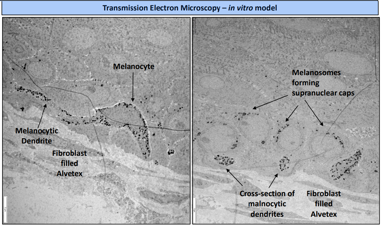
Creating a truly accurate human tissue cannot be rushed. Unlike other commercially available artificial skin, REPROSKIN™ is entirely comprised of primary cells, which secrete their own endogenous extracellular matrix (ECM). The inclusion of additional cell types, such as melanocytes in our pigmented skin models, enables studies of specific functions. 

The anatomical accuracy of REPROSKIN™ has been extensively characterised. Using our longstanding experience in human fresh skin, we have extensively characterised numerous markers of skin biology in both REPROSKIN™ and fresh skin, creating robust and reproducible engineered skin equivalents.

Our custom-manufactured plasticware enables a wide variety of tests and applications, including the safety or efficacy of pharmaceuticals, cosmetics, chemicals or even devices that interact with the skin. Measurements, such as changes in protein composition and biomarker expression, barrier function, water loss, or the release of inflammatory mediators, are all possible, delivered in our labs by our team of expert scientists. 

Alvetex Scaffold skin model

REPROSKIN: biologically accurate engineered human skin

Expert lab service for the testing of pharmaceuticals, chemicals, cosmetics or devices on living human skin

REPROCELL offers an exciting and novel solution for the study of live human skin in support of pharmaceutical, cosmetic, chemical and consumer product markets. Tests are conducted at REPROCELL's facilities on a fee-for-service basis; our scientists will work with you to create a customised proposal that meets your research objectives. 

REPROSKIN™ is comprised of epidermal keratinocytes and dermal fibroblasts, with a morphology comparable to native skin. In addition, we can introduce additional cell types into REPROSKIN™, creating customised skin models for specific applications:  

  • Wound healing model: the resolution of a controlled wound to the epidermis and/or dermis is monitored over a number of days to assess the wound healing properties of drug compounds, lotions, creams or devices.
  • Aging model: cells from adults over 50 years of age are included to reproduce aspects of aged skin.
  • Pigmented skin model: melanocytes are included in the 3D full thickness skin to create pigmented skin representative of different skin tones. The effects of UV exposure, in the presence or absence of protective creams or lotions, can be assessed. 
  • Sensory skin model: sensory neurons are included within the skin to develop assays for testing products that may evoke a sensorial response.
  • Immune competent skin model: includes immune cells for modelling inflammatory responses

Key features of the REPROSKIN™ system include:

  • Easy access to the epidermal surface while the skin remains alive in culture, allowing the application of topically applied test substances, devices or measurement probes.
  • Innovative tissue seal between upper and lower compartments of the culture system, minimising leakage of substances when applied to the epidermal surface (no “edge-effect”).
  • Compatible with various functional measurements that can be easily made non-invasively within the system (e.g. pH, TEWL (tran, TEER and dye penetration).
  • The well inserts containing the live skin models can easily be transferred to different plate formats without disruption, increasing the flexibility of the test systems.
  • The skin models are compatible with the ability to generate wounds (cuts/scratches/burns) in a controlled manner and then to observe and measure wound healing over time.
  • With easy access to the skin surface, the skin can be exfoliated, for example via tape stripping methods.
  • Inserts and plates are compatible with UV instruments, such as BioSun Irradiator, for the delivery of fixed amounts of UVA and UVB radiation, resulting in skin darkening and assessment of sunscreen products.
  • Our skin models are compatible with the majority of endpoint assays that can be performed on biopsied human skin (e.g. histology, imaging, protein/gene expression, etc.).
  • Instrumentation currently used in the clinic to monitor skin parameters are directly compatible for use in our in vitro models – closing the gap between the laboratory and clinical trial – hence increasing relevance and predictive accuracy of our in vitro tests and potentially reducing costs and time of clinical trials and even aiding their design.
Wound Healing and Skin Damage Models

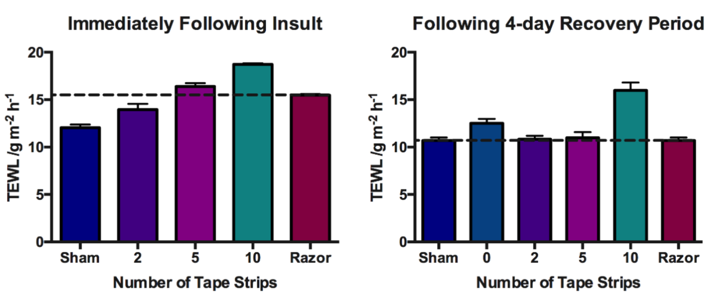
The REPROSKIN™ models allow for the controlled generation of wounds, such as cuts, scratches and burns, enabling the observation and measurement of wound healing over time. Additionally, these models provide easy access to the skin surface for exfoliation, using methods like tape stripping or the application of devices. This allows for the assessment of structural or functional changes in the skin following the application of a test substance or device.

A cut is created in the epidermis alone

A cut is created in the epidermis alone, or in both the  epidermis and dermis, in a controlled manner, and the wound healing process is monitored for up to 14 days.

Trans-epidermal water loss (TEWL) increases immediately post-insult

Trans-epidermal water loss (TEWL) increases immediately post-insult, indicating barrier disruption.

The wound has closed and a new stratum corneum is present after 3 days of culture

The wound has closed and a new stratum corneum is present after 3 days of culture, post-insult.

Aging Skin Model

The aging skin model has been developed using keratinocytes and fibroblasts from the donors over the age of 50. This innovative approach aims to replicate aspects of aging skin, displaying epidermal thinning and diminished extracellular matrix production in the dermis. By mirroring age-related changes in skin phenotype, this model is a valuable tool for testing cosmetic products that enhance the appearance of the skin. For example, in-depth exploration of glyco-repair mechanisms or the effects of retinoids and small molecules, can readily be conducted. 

Contact REPROCELL today to discuss a customised approach to testing your skin aging products.

skin model

Pigmented Skin Model

REPROCELL employs a comprehensive approach in crafting pigmented human skin models, intricately designed to simulate the complexities found in real skin. This involves integrating melanocytes, specialized cells responsible for melanin production. The formation of supra-nuclear caps, structures found in the skin's epidermis, adds further authenticity to these models. Notably, these models demonstrate evidence of tanning post-UV exposure, showing their responsiveness to environmental stimuli. Furthermore, they exhibit the protective properties of melanin against harmful UV rays, substantiating their use in understanding skin defence mechanisms. By representing various ethnicities, these models cater to the diverse range of human skin tones, making them invaluable in developing inclusive skincare products. The application allows investigation of both whitening and darkening products, as well as UV resistance formulations, providing a robust platform for the progression of skincare research and development.

skin pigment
skin model 3

Key Application in SPF 

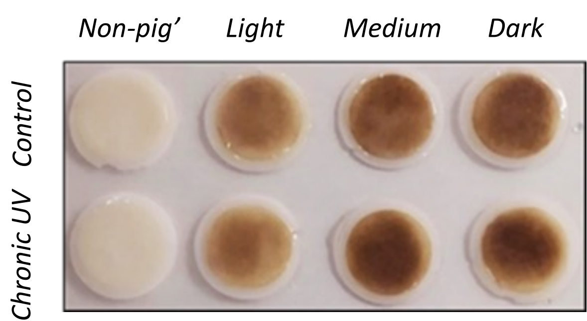
Pigmented models acts as a platform for testing sunscreen formulations. This model provides the leading method of testing topical application of SPF (sun protection factor) products to the surface of the human skin.

REPROCELL tested both the pigmented and non-pigmented model in chronic UV light
REPROCELL tested both the pigmented and non-pigmented model in chronic UV light to determine the result on the skin in the absence of sunscreen. As expected, the skin models were damaged by UV exposure, containing characteristic sunburn cells with a detached epidermis and blistering. In the presence of sunscreen with SPF there was no visible damage, with models presenting with a well formed and organised epidermis.

Diffuse Barrier/Skin Damage Model

The core objective of every skincare product is to safeguard the skin from damage and ensure a positive user experience. REPROCELL has developed a tape stripping technique utilizing bioengineered models to simulate various types of damage to the epidermal layer. Through rigorous testing, this innovative skin model demonstrates the ability to recover from surface damage after topical applications such as moisturizers. 

tape strip

Testing your skin products 

Does your razor prevent epithelial damage? Does your new formula gel enable a smoother shave? How do the layers of the skin repair after using your product?

Trans-epidermal water loss (TEWL) is useful for assessing barrier function, which can indicate skin damage caused by various factors, including chemicals, physical insults, or pathological conditions such as atopic dermatitis. These are some of the more common applications, but the model is useful for any type of skin damage. The rates of TEWL increase in proportion to the level of damage.

Immune Competent Skin Model

Pmmune competent skin momdelOur Immune Competent Skin Model is a sophisticated representation that mimics the intricate immune network within the skin. The model encompasses both the epidermis and dermis, where Langerhans cells, dendritic cells, and dermal macrophages collaborate harmoniously to fortify the skin's defense.

To develop an in vitro skin model containing Langerhans cells, the human cell line MUTZ-3 was integrated into the already established full-thickness models by co-seeding with HEKn. These MUTZ-3-derived Langerhans cells show expression of CD1a and langerin in the epidermis. This unique model provides a comprehensive understanding of the immune cells residing in the skin, offering researchers and scientists a valuable tool for in-depth exploration and the study of skin immunology.

 Full thickness models containing MUTZ-3-derived Langerhans cells show lower homeostatic levels of inflammatory cytokines, but show an increased production of these mediators in response to allergens, irritants and particulate matter exposure.

Contact our scientists today to investigate your compounds in our immuno-competent engineered skin or fresh skin tissues.


Make an inquiry