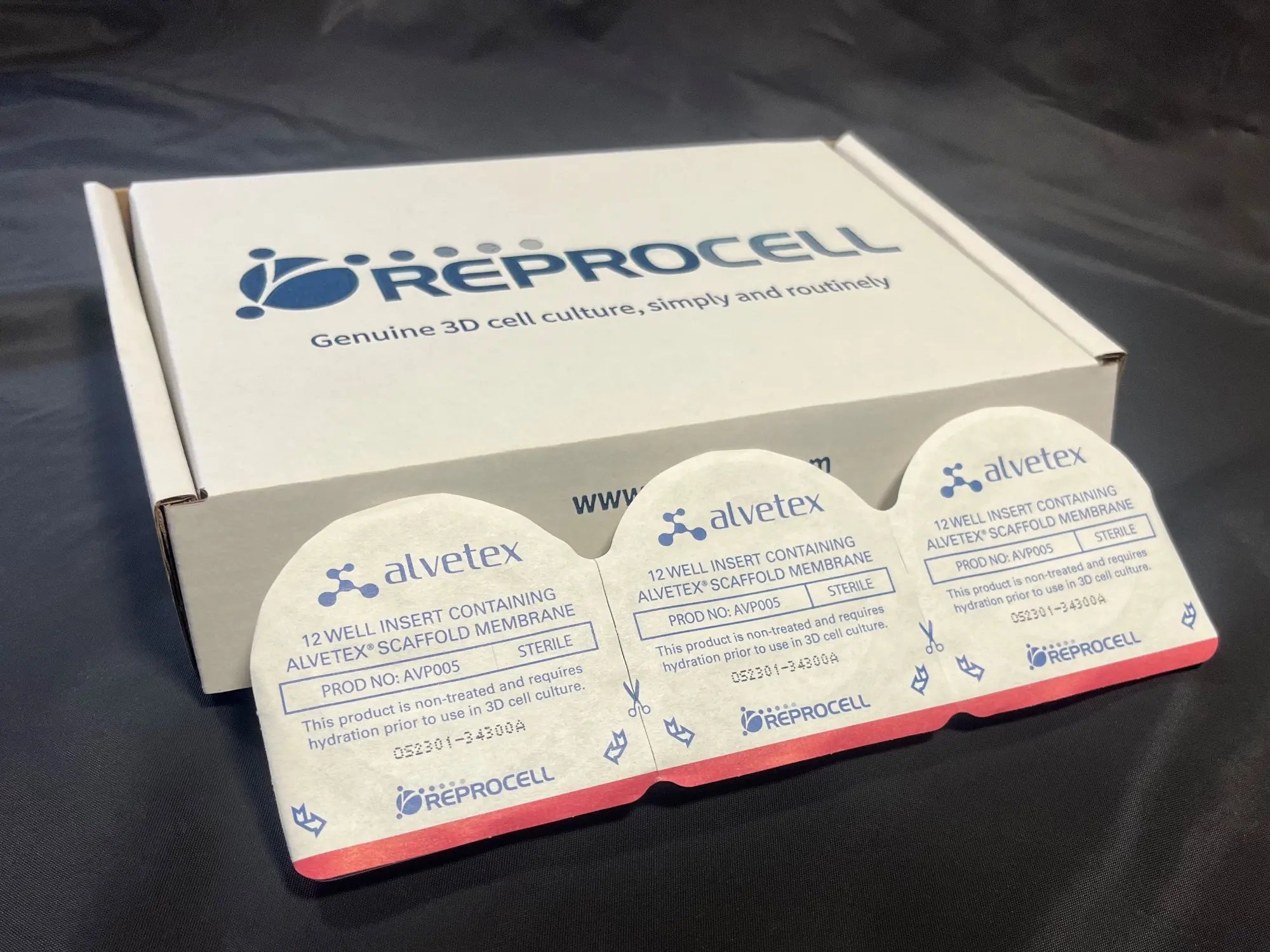
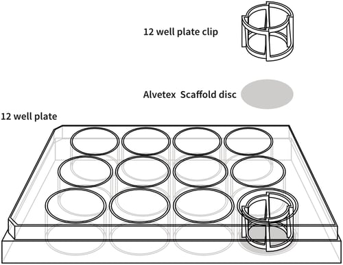
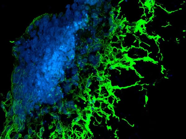
A range of formats for all your applications
Alvetex Scaffold is primarily designed for three dimensional culture of dissociated mammalian cells within the scaffold, forming three dimensional associations as they propagate and migrate.


Above: Scanning electron microscope image of Alvetex Scaffold, to highlight its porous structure. A: a 200 µm thick Alvetex Scaffold disc. B: Close up of Alvetex Scaffold voids with dimensions of approximately 42 µm in diameter and interconnects of approximately 13 µm in diameter.
Alvetex Strata is primarily designed to support the growth of cells and intact tissues on the surface of the membrane.


Above: Scanning electron microscope image of Alvetex Strata. C: a 200 µm thick Alvetex Strata disc. D: Close up of Alvetex Strata voids with dimensions of approximately 15 µm in diameter and interconnects of approximately 5 µm in diameter.
Choosing the right Alvetex format based on assay type
Suggested guidelines for the use of Alvetex formats for cell applications and assays.
+++ = most suitable
++ = suitable
+ = least suitable
n/a = not applicable
Ranking is based on Alvetex disc format suitability, the likely cell yields and therefore signal generation, and whether exogenously added chemicals and/or cells can be contained to only one side of the membrane.
| Types of assay | Alvetex Scaffold 6 well insert |
Alvetex Scaffold 12 well insert |
Alvetex Scaffold 24 well insert |
Alvetex Scaffold 12 well plate |
Alvetex Scaffold 24 well plate |
Alvetex Scaffold 96 well plate |
Alvetex Strata 6 well insert |
Alvetex Strata 12 well insert |
|---|---|---|---|---|---|---|---|---|
| Viability/Proliferation/ Metabolic Activity Assays | +++ | +++ | +++ | +++ | +++ | +++ | +++ | +++ |
| Toxicity Assays | +++ | +++ | +++ | +++ | +++ | +++ | +++ | +++ |
| Gene Expression assays (qPCR/ microarray) | +++ | +++ | +++ | +++ | +++ | +++ | +++ | +++ |
| Protein Expression assays (e.g. western blot) | +++ | +++ | +++ | +++ | +++ | +++ | +++ | +++ |
| Air-liquid Interface assays | +++ | +++ | +++ | N/a | n/a | n/a | +++ | +++ |
| Cell Signaling assays | +++ | +++ | +++ | +++ | +++ | +++ | +++ | +++ |
| Permeability assays | +++ | +++ | +++ | n/a | n/a | n/a | +++ | +++ |
| Transfection assays | +++ | +++ | +++ | + | + | + | +++ | +++ |
| Co-culture assays | +++ | +++ | +++ | ++ | ++ | ++ | +++C | +++C |
| Invasion assays | +++ | +++ | +++ | + | + | + | ++C | ++C |
| Migration assays | +++ | +++ | +++ | + | + | + | ++C | ++C |
| Histology | +++ | +++ | +++ | ++ | ++ | ++ | +++ | +++ |
| Immunostaining (IHC/IF) | +++ | +++ | +++ | ++ | ++ | ++ | +++ | +++ |
| Confocal microscopy | +++ | +++ | +++ | ++ | ++ | ++ | ++ | ++ |
| Live cell imagingA | +++ | +++ | +++ | ++ | ++ | ++ | ++ | ++ |
| Ex vivo tissue maintenance | +++ | +++ | +++ | ++ | ++ | ++ | +++ | +++ |
| Live cell retrievalB | +++ | +++ | +++ | ++ | ++ | ++ | ++ | ++ |
Notes:
A: The growth of cells cannot be followed by traditional light microscopy as in 2D, but as with ex vivo tissues, 3D structures have to be evaluated using histology or confocal microscopy. Alternatively cell proliferation can be monitored using a viability assay such as the MTT.
B: The exact number of cells retrieved from Alvetex varies with the invasiveness of the cell line cultured, e.g. epithelial vs. fibroblastic. Although the three-dimensional structure of Alvetex precludes all 100% of the cells from being routinely retrieved, calls can be retrieved in adequate numbers for quantitative down-stream processes, e.g. flow cytometry.
C: When designing co-culture, invasion or migration set-ups for Alvetex Strata, please keep in mind that some cell lines (e.g. epithelial) have a tendency to multi-layer on top of the substrate rather then invade into it.


