Frequently Asked Questions about Alvetex® 3D Cell Culture
● Download the PDF version of these FAQs
Here are some frequently asked questions concerning 3D cell culture on Alvetex with our answers. If you can’t find the help you need here, please contact us.
Format and Supply
- How is Alvetex supplied?
Both Alvetex Scaffold and Alvetex Strata products are gamma irradiated and supplied in sterile, individually-wrapped blister packs. - What formats of Alvetex are available?
Alvetex Scaffold is currently available in the following formats: 12 well plates (AVP002), 24 well plates (AVP006), 96 well plates (AVP009), 384 well plates (AVP010), 6 well inserts (AVP004) and 12 well inserts (AVP005). Alvetex Strata is available in the 6 well inserts (STP004) and 12 well inserts (STP005) formats. The well inserts fit a range of culture plates from different manufacturers as well as REPROCELL’s Well insert holder in a deep Petri dish (AVP015). For more information, please see the REPROCELL Brand: Alvetex on our website or our Alvetex 3D Cell Culture Systems products page. - Are other formats available?
We are continually optimising our current products and developing new formats. Custom-made formats of Alvetex are available for collaborations, please contact us for details. - What are the dimensions of the of Alvetex discs?
The following numbers are the same for both Alvetex Scaffold and Alvetex Strata discs:
| 12 well plates AVP002 / STP002 and 6 well inserts AVP004 / STP004 |
24 well plates AVP006 / STP006 and 12 well inserts AVP005 / STP005 |
24 well inserts AVP012 |
|
| Diameter | 22.0 mm | 15.0 mm | 11.5 mm |
| Available diameter inside insert |
18.5 mm | 12.3 mm | 8.5 mm |
| Growth area | 269.0 mm2 | 118.9 mm2 | 56.8 mm2 |
- What is the difference between plates and inserts?
In the plates (12-well and 24-well formats), the Alvetex membrane is at the bottom of the well so that access to the medium is from above the membrane only. In the insert, the membrane is suspended so that access to the medium is from both above and under the membrane. This is why inserts are recommended for culture periods of more than 7 days. Also in the insert, the bottom of the well (into which the insert is suspended) is free of cells, allowing for paracrine co-culture setups. - It seems from looking at website that the inserts are the most suitable for most of applications (i.e. histology, co-culture...) Why?
Because inserts get access of medium to the bottom of the membrane, they can support growth of cells for longer periods that the plate format. We recommend using plate formats for shorter culture period (up to 7 days) and inserts for longer culture periods (more than 7 days). For smaller-size membranes (i.e. higher throughput), however, some multi-well plate formats (24 wells and 96 wells) are currently only available as plates, not as inserts. - Can Alvetex plates be used for co-cultures?
Two co-culture set-ups are achievable using plate formats. Firstly, by mixing two cells populations and growing them as an heterogenous co-culture in the membrane at the bottom of the well. Secondly, by combining a well bottom membrane and an insert in the same well, for a 3D-3D co-culture. Please note that, as plates are recommended for culture periods up to 7 days only, all co-culture setups are best performed using insert formats as they provide a more even nutrition through the depth of the membrane and sustain cell growth for longer periods. 3D-3D co-culture can be achieved with inserts if placed within a holder for deep petri dishes (AVP015). - What is the difference between Alvetex Scaffold and Alvetex Strata?
The void size of Alvetex Scaffold is greater than the void size of Alvetex Strata. Functionally, this results in some epithelial-type cell lines forming a multilayer on top of Alvetex Strata rather than migrating into it, as would be observed when the same cells are cultured in Alvetex Scaffold. By comparison, more invasive or fibroblast-type cell lines will penetrate into both Alvetex Strata and Alvetex Scaffold. For advice as to which void size is best suited to your specific project, please contact technical support. - Is Alvetex supplied sterile?
Alvetex products have been sterilised by gamma irradiation and remain sterile until the blister packs are opened. - Is Alvetex reusable?
Alvetex is a single use, disposable product. - Does Alvetex have an expiry date?
There is no expiry or shelf life for Alvetex. The plate or inserts are sterile and ready for use as long as the sterile packaging seal remains intact.
Chemical and Physical Properties
- What is Alvetex made from?
Alvetex is a cross-linked polystyrene scaffold supplied with a thickness of 200 µm. It is highly porous, inert and does not degrade. - What are the void sizes and interconnect within Alvetex?
Alvetex Scaffold voids have an average diameter of 40 µm with interconnects of approximately 13 µm in diameter. Alvetex Strata voids have an average diameter of 15 µm with interconnects of approximately 5 µm in diameter. - What is the porosity of Alvetex?
Both Alvetex Scaffold and Alvetex Strata have a porosity of at least 90%. - What is the stiffness of Alvetex?
We do not perform any mechanical evaluation of Alvetex, although a Young’s modulus value of 77 kPa has been recorded for Alvetex Scaffold in the literature (Drug Discovery Today, 2013, 18(11-12):533-540). As Alvetex is at least 90% porous, the mechanical environment experienced by cells within Alvetex is likely to be more dependent on cell-cell contact than on cell-substrate contact. The potential contribution of any coating material used would also need to be considered. - Can you autoclave Alvetex?
The Alvetex discs themselves can be autoclaved, ensuring they have been removed from the insert or plate first. - Is Alvetex chemically resistant?
Owing to the cross-linked nature of the polystyrene substrate, it is chemically resistant; however, the surface properties can be affected by certain chemicals. Some solvents like acetone may swell the substrate. Please contact technical support if you have a query about a specific chemical. - Is there disc to disc variability?
Each batch of Alvetex is tested for its physical properties, porosity and suitability for cell attachment and viability. Only batches fulfilling strict criteria are released by our ISO9001:2008 certified Quality System.
Culturing your Cells on Alvetex
- Which cell types have been tested on Alvetex?
We have released many protocols for culturing the following cell types on Alvetex Scaffold: 3T3, HaCaT, HepG2, TERA2.cl.SP12, CHO-K1, LN229, full thickness skin equivalent, SW480, SW620, PC3, BT474, MG63, primary rat MSCs, MCF-7, Caco-2 and the co-culture of Caco-2 with CCD-18co cells, which can be accessed through the Alvetex Protocols page of the website. Most of the cell lines included in our Alvetex Protocols page have also been cultured on Alvetex Strata. Other cells which have been successfully grown on Alvetex Scaffold include H1299, U118-MG glioblastoma cells, bone marrow stromal cells, primary hepatocytes, Upcyte hepatocytes, neurospheres, adipose tissue-derived stem cells, human pluripotent stem cell-derived neurons, cylindroma primary cells, MET4 squamous carcinoma cells, L929 mouse fibroblasts, neural crest cells, human nucleus pulposus cells, chondrocytes, primary chick embryonic tissue, brain tissue slices, equine oviduct cells, spermatogonial stem cells, human disc cells, melanocytes, A375/A2058 melanoma cells, 3T3-L1 cells, prostate cancer cells and prostate stem cells, H9 human embryonic stem cells, CRL-11372 osteoblasts, HEK293 human embryonic kidney cells, JJ012 chondrosarcoma cells, NG108 stem cells, MBT-2, MB49 mouse bladder cancer cells, rat primary urothelial cells, primary retinal pigment epithelium, urothelial cells, myofibroblasts, astrocytes, primary and secondary ESC-derived hepatic cells, primary keratinocytes, pancreatic duct and stromal cells, GSC glioblastoma stem cells, b.END-3 endothelial cells, HCT116 cells, among others. - Do you have to use all the discs in an Alvetex plate at the same time?
For 12-well and 24-well plate formats, discs and holders can be removed under the hood (i.e. under sterile conditions) and transferred to another plate for later use. For the 96-well and 384-well plate formats, Alvetex is heat-welded at the bottom of the plate and cannot be removed, so that a full plate of Alvetex will have to be used at any one time. - Can Alvetex be coated?
Yes, most standard coating methods for cell culture plastic are compatible with Alvetex Scaffold including the use of extra-cellular matrix proteins, poly D/L lysine etc. In addition, a thin layer of collagen gel can be cast on top of Alvetex to promote culture of a monolayer of epithelial cells. A range of protocols are available in our Alvetex Protocols - What is the significance of ethanol-treating Alvetex?
Alvetex needs to be treated with ethanol in order to render it hydrophilic. If this step is missed out and cells are seeded directly onto the untreated scaffold, they sit in a droplet on the top and are unable to penetrate and grow in 3D. Once Alvetex has been treated with ethanol, it must be kept hydrated with either PBS or cell culture medium to maintain it into an hydrophilic state. - How long can you culture cells for in different Alvetex formats?
Depending on the cell type and the method of analysis to be performed, Alvetex 12 well plate or 24 well plate formats are usually recommended for cell culture experiments lasting between 1 and 7 days. Well insert formats are more suited to long term experiments of between 1 and 5 weeks. For experiments lasting more than 2 weeks, we recommend suspending inserts in deep petri dishes using our dedicated petri dish holder (AVP015), so that cultures can benefit from a larger volume of medium. - What happens if less than the recommended number of cells is seeded onto Alvetex?
If cells are usually kept at high density in 2D, seeding them at less than the recommended cell number could hamper their viability and/or growth. If cells can be kept at lower density in 2D, seeding at lower than recommended density will delay them in reaching confluence in Alvetex so longer time points might be needed for analysis. - What happens if more than the recommended number of cells is seeded onto Alvetex?
The seeding densities recommended are guidelines and may need to be optimised depending on cell type. If too many cells are seeded, the substrate may become saturated, and cells may migrate out and grow in 2D on the surrounding plastic-ware. We recommend transferring inserts to a fresh plate the day following cell seeding to prevent interference from 2D-adherent cells. - What happens if cells are seeded in a larger volume than is recommended?
If larger cell seeding volumes are used then the cells may get drawn through the scaffold (concentrated seeding method) or over the insert windows (disperse seeding method) before they are able to attach to Alvetex, leading to cells adhering to the plastic ware below. We recommend transferring inserts to a fresh plate the day following cell seeding to prevent interference from 2D-adherent cells. - What does it mean for Alvetex inserts that 3 positions are available?
This can refer either to the medium level or to the deep petri dish holder (AVP015). For inserts in individual wells, the medium level can be adjusted to be just touching the bottom of the membrane (air-liquid interface), above the membrane but under the windows in the insert wall (above and below separately) or above both the membrane and the windows in the insert wall (submerged). Which level to use depends on the cell type and application of the culture (e.g. airway epithelium differentiation). In the deep petri dish insert holder (AVP015), each holding position is manufactured like a ladder with three possible heights at which the insert can be positioned. This allows one insert to be at one height and another insert in the same dish to be at another height, which can be useful if distinct cell populations with different feeding preference are co-cultured in the same dish (i.e. 3D-3D paracrine co-culture setup). - What is meant by “independent compartments enable 3D growth with two different media constituents”?
The ‘above and below’ medium level is one where there is medium above the membrane but at a level below the windows in the insert wall. If there is a confluent mass (or layer) of cells in the insert, and specific small molecules are added to the medium either on the inside or the outside (not both) of the insert, then the side of the cells exposed to the inside of the insert will experience different conditions than the side of the cells exposed to the outside of the insert. If the cells are not confluent in the insert, then the gradient will equilibrate quickly over time. Also, the volume of medium inside the insert will be small and will need renewing frequently to keep providing sufficient nutrition to the cells at the top of the substrate. - What is the difference between the second and third medium level position?
In the third position, the medium level comes above the windows in the insert, so that there is communication between the inside and outside of the insert independently from the state of confluency of the cells and the nutrition provided is maximal. This is recommended to maximise growth or for cells with higher metabolic needs. - What is the minimum required volume of culture media in a well when using Alvetex well inserts?
In a standard 6-well plate, both 6-well inserts and 12-well inserts are suspended 4.7 mm above the floor of the well. Plate wells are 34 mm wide. Therefore, a minimum of 4.4 mL (approx.) liquid is required to reach the Alvetex disc in the insert. In a standard 12-well plate, 12-well inserts are suspended 4.7 mm above the floor of the well. Plate wells are 22 mm wide. Therefore, a minimum of 1.8 mL (approx.) liquid is required to reach the Alvetex disc in the insert. If in doubt, users can manually check the exact volume needed for their chosen feeding option with PBS first (which is inexpensive, is used for preparing Alvetex before cell seeding and can be used to keep Alvetex hydrated until the medium and cells are ready). - For plate formats, do you have to remove the disc holders before seeding the cells?
No, the disc holders are intended to be kept in place at all times during culture. The area covered by the holders does not significantly affect cell growth, due to the much larger area available for cell attachment and growth within the depth of the scaffold. - What happens if air bubbles are present underneath the insert?
Air bubbles underneath inserts should be avoided, as they will create areas where nutrients and waste exchange is not occurring properly. To avoid air bubbles, gently fill the well while holding it at an angle until a visual check can be done that there are no bubbles underneath the insert. If an air bubble is present, the insert can be lifted off and gently placed down again at angle to try and get rid of the air bubble. - What tissue culture models are achievable with Alvetex?
Alvetex can be used to produce a range of in vitro tissue culture models. The simplest model is to culture a single cell type in each well. With insert formats, it becomes possible to co-culture several cell types, either separately to test for the effect of soluble factors or in close proximity to test for tissue invasion. The full range of co-culture set-ups achievable with Alvetex insert formats is detailed in our Applications Notes page. Even more complex models, whereby several cell types are seeded sequentially in Alvetex to approximate the architecture and functionality of in vivo tissue, can also be obtained. Skin models are such an example and a detailed protocol is available in our Alvetex Protocols - Can you perform co-culture experiments in Alvetex?
There are several options available; these are described in detail in the co-culture application note, which is available in the Alvetex Application Notes and Whitepapers page of our website. - Can a layer of 2D cell seeded in the well of the plate be co-cultured with 3D cells growing in the Alvetex disc on the plate clip above?
This is possible using the insert formats where 2D cells can be grown at the bottom of a conventional plate, and then 3D cells seeded into an insert can be added to that well with the 2D cells already at the bottom. We recommend seeding the 3D cells in a separate well first and leave them there for at least 48 hours to attach to the membrane before transferring the insert to the well with the 2D cells, to avoid cells seeded in 3D detaching soon after seeding and ending up at the bottom of the well. - Are complex tissue-like models achievable with Alvetex?
Cultures using relevant cell types seeded and grown sequentially in Alvetex can approximate the architecture and functionality of in vivo tissue, with a view to produce improved in vitro assays without the limitations of in vivo and ex vivo assays, i.e. species-specificity and tissue degradation, respectively. Skin models are such an example for which a detailed protocol is available in our Alvetex Protocols page. For other complex models developed by Alvetex users, please consult our Alvetex Publications page. - Is Alvetex suitable for cell invasion and cell migration assays?
An application note describing cancer cell invasion and migration within Alvetex Scaffold can be found in the Alvetex Application Notes and Whitepapers page of our website. Please note that, due to smaller void size, some epithelial-like cell types might only show very limited migration/invasion into Alvetex Strata, and will instead tend to monolayer or multilayer on top of Alvetex Strata. - Is Alvetex compatible with in vivo transplantation?
Alvetex is not approved for use with in vivo - Can you explant tissues onto Alvetex?
It is possible to take explanted tissue and maintain it onto Alvetex, and well-insert formats are recommended for this purpose. For short-term studies where cell migration/expansion into the substrate is not required (e.g. live cell imaging of ex vivo cells in freshly explanted tissue), Alvetex Strata is recommended. For long-term experiments where cell migration/expansion in the 3D substrate is actively desired, both Alvetex Strata and Alvetex Scaffold can be used, keeping in mind that larger/less invasive cells will show reduced penetration in Alvetex Strata compared to Alvetex Scaffold due to differences in void size. Detailed protocols for both methods are available in our Alvetex Application Notes and Whitepapers page.
Analyzing your Alvetex Cultures
- Are there any protocols available for users?
A range of example protocols can be found in the Alvetex Protocols page of our website. These are being continually updated and expanded upon and currently include general information about choosing and using Alvetex formats; examples of how to grow specific cell types; compatible analytical techniques and specialised applications. - How do you remove Alvetex discs for analysis?
In plate formats, each Alvetex disc is held at the bottom of the well by a holder which can be lifted using a pair of flat-ended forceps. The Alvetex disc itself can then be retrieved by gently lifting it at its edge using a pair of fine-ended forceps, then gently but firmly taking hold of the disc’s edge with a pair of flat-ended forceps and lifting it out of the well. In well-insert formats, each Alvetex disc is held between the two-part base and body of the insert. The base can be unclipped from the body by using gloved fingers or a pair of flat-ended forceps to undo the clips at the base of each of the three windows in the body of the inserts. The Alvetex disc can then be gently lifted off and held using flat-ended forceps. A protocol detailing this procedure is included in our Alvetex Protocols - How can you see your cells growing in 3D in Alvetex?
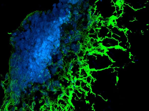
Live cells cultured in Alvetex can be visualised with a range of methods, each of which is described in detail in the protocols available from our Alvetex Protocols For a quick check of cell population confluency or general cell morphology, dyes such as neutral red can be used with conventional light microscopy. For continuous imaging of live cells over a few hours to several days, live cell imaging with fluorophores and confocal microscopy can be used. - Does Alvetex autofluoresce?
Alvetex does not autofluoresce at wavelengths used for the detection of common fluorophores and at standard laser power settings (i.e. below excessive laser power that produces autofluorescence in all elements within the sample). When viewed under a conventional microscope, light refraction through the thickness of Alvetex will result in background which can be misinterpreted as autofluorescence. This issue is easily resolved using a confocal microscope. Alvetex should be blocked and washed thoroughly as described in the dedicated protocols available from our Alvetex Protocols page to avoid non-specific binding of fluorophores. It should be noted that as Alvetex is hydrophobic, lipophilic stains will bind strongly to it, therefore working concentrations of lipophilic dyes must be carefully optimised or alternatives should be sought to avoid double-staining. - Can you visualize live cells using Alvetex?
For following culture progress, the use of dyes can be employed to visualise cells against the scaffold background using light microscopy. A full protocol for this simple method is available within the Alvetex Protocols page of our website. More complex techniques that are commonly used for tissue processing can also be implemented with excellent results. - What analysis techniques are compatible with Alvetex?
Alvetex is compatible with a broad range of general cell and molecular techniques. These include various cell viability assays, imaging techniques (fixation, embedding, sectioning, and staining for histology, immunocytochemistry, electron microscopy), confocal and live cell microscopy, cell transfection, cell retrieval, protein, and nucleic acid extraction. Please visit our Alvetex Protocols page for the full range of available protocols. - Is it possible to cut Alvetex before processing it for analysis?
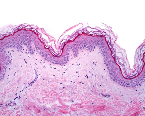
Cutting Alvetex into several pieces (e.g. so that each piece can be used for a different analytic method) can be done with either a sharp razor blade or fine scissors. Cutting Alvetex into thin sections requires embedding in either paraffin wax or OCT, depending on the final application. - What histological clearing agent do you recommend for use in the preparation of sections from cells grown in Alvetex Scaffold?
We recommend the use of Histo-Clear as part of our tissue processing protocol for histological staining. Histo-Clear is much safer to use than xylene (removing the need to store, use and dispose of xylene) but also improves results. Alvetex is a cross-linked form of polystyrene and on the whole is resistant to xylene and it is possible to use this reagent. However, slight swelling of the scaffold material may occur after exposure to xylene and this may consequently result in section/ cell loss unless the samples are handled carefully. Histo-Clear leaves tissue less hard and brittle than xylene, facilitating the cutting of thin sections and prolonging microtome blade life. Nuclear morphology is rendered in fine detail. Histo-Clear enhances the clarity and vibrance of acidophilic stains and improves staining of Harris’ Hematoxylin with a brighter Eosin background. - Can you remove cells from Alvetex?
Partial cell recovery is possible and is greater from Alvetex Scaffold than from Alvetex Strata due to differences in void size, especially for highly invasive cells. Cells can be removed from the substrate using a combination of trypsin and mild agitation. For techniques requiring extraction of material from the substrate, such as total protein or nucleic acid isolation, cell retrieval is not required or recommended. For example, protocols of cell retrieval and protein/nucleic acid extraction, please visit our Alvetex Protocols - How do you estimate cell confluency in Alvetex?
Simple dyes can be used to estimate cell culture confluence and viability. A full protocol for a simple method is available in the Alvetex Protocols page of our website. An extensive number of end-point visualisation techniques are also compatible, including live cell imaging, fluorescent marker analysis, confocal analysis, biochemical assays, histological analysis using a range of cytological stains and electron microscopy. - Is it possible to cryosection Alvetex?
A protocol is available in the Alvetex Protocols page of our website. - Can you perform FACs analysis on cells from Alvetex?
Cells removed from Alvetex can be used for FACS analysis. Please refer to our Alvetex Protocols page for details on how to retrieve cells from Alvetex. - Is there a protocol for performing transfection in Alvetex?
Yes — a transfection kit is commercially available and is produced in conjunction with our partner company, Mirus Bio. A link his provided in our Alvetex Protocols - What can I do if my paraffin-embedded section of Alvetex does not adhere to the microscope slide?
Poor adhesion can be the result of having limited cell numbers within Alvetex, of having used an inadequate microscope slide, or of excessive mechanical disruption during staining. To address these issues, we recommend either increasing the cell culture incubation time to attain a higher density of cells before processing, using electrostatic charged slides (e.g. Superfrost™ plus), and/or using gentle manual staining rather than automated staining. - Can you get reproducible data using Alvetex?
Reproducibility of data using Alvetex is very good. Where the same cell suspension has been used, the same growth pattern for the cells is consistently obtained. It should be noted that some cell types prefer to occupy the top quarter of the scaffold, whereas others invade all the way through.
Evidence-Based Technology
- Have any papers been published regarding Alvetex?
Numerous peer-reviewed scientific articles describe the development and application of Alvetex 3D cell culture, which can be accessed from the Alvetex Publications Scientists at REPROCELL are continuously researching novel applications for this technology and are publishing and presenting data when appropriate.