Example Protocol for the Culture of the Human Colon Fibroblastic CCD-18co Cell Line on Alvetex® Scaffold in Well Insert and Well Plate Formats
● Download this protocol as a PDF (1.7 MB)
1. Introduction
CCD-18co is a human fibroblast cell line isolated from normal colon tissue. Being a non- transformed cell line, CCD-18co is a popular choice as a normal control in cancer studies[1,2] and also to form a fibroblastic compartment in co-culture models of epithelial-stromal interactions in colon cancer[3]. In their own right, CCD-18co cells are also used to investigate mechanisms of pathological fibrosis following colon inflammation[4,5].
Alvetex Scaffold is available in several cell culture formats including 24 well plate (AVP006), 12 well plate (AVP002), 6 well insert (AVP004), 12 well insert (AVP005), and 24 well insert (AVP012).
24 well and 12 well plates are suitable for shorter term cultures and for applications where limited cell penetration into the scaffold is required. Well insert formats generally support longer term cultures and deeper cell penetration into the scaffold. They also provide for conveniently tailored media set ups (see the Alvetex Scaffold Quick Start protocol).
The availability of two different well insert formats enables choice on the basis of desired culture size and cell expenditure. 6 well inserts can be placed in conventional 6 well plates, while 12 well inserts can be placed in either 6 well plates or 12 well plates, depending on media requirements. Alternatively, both insert types can be housed in the dedicated Well Insert Holder in Deep Well Petri Dish (AVP015) to allow for increased media volumes and prolonged cell culture.
2. Methods
2.1. Preparation for 3D Cell Culture on Alvetex Scaffold
- CCD-18co cells (ATCC, CRL-1 459) were routinely maintained in T-75 flasks.
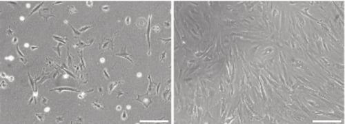
Figure 1. Phase contrast micrograph of CCD-18co cells grown in conventional 2D culture plates. Image shows cells at low (left) and high (right) confluency. Scale bars: 200 µm.
- Complete growth media consisted of: high glucose (4.5 g/L) DMEM supplemented with 10 % v/v FBS, 2 μm L-glutamine, 100 U/mL Penicillin/Streptomycin and 0.1 mM non-essential amino acids.
- Cells were harvested by trypsinisation and centrifuged for 5 minutes (1000 rpm). The supernatant was discarded and the cell pellet was re-suspended in an appropriate volume of media for cell counting by Trypan Blue.
- Cells were re-suspended at a concentration of 4.0 × 10⁶ cells/mL for seeding.
2.2. 24 well Plate Format (AVP006)
- Alvetex Scaffold 24 well plates were prepared for seeding with a 70 % ethanol wash (2 mL per well) and subsequent media washes (twice with 2 mL of media each).
- 500 μL of the cell suspension was added to the centre of the Alvetex Scaffold disc, which was equivalent to 2 × 10⁶ cells per well.
- The plate was incubated for at least 1 hour at 37 °C with 5 % CO2 to allow the cells to settle into the scaffold.
- 1.5 mL of media was added to each well taking care not to dislodge cells from the Alvetex Scaffold.
- Plates were re-incubated and maintained by complete media exchange every other day.
Note: This method can be applied to the use of Alvetex Scaffold in 12 well plate format (AVP002). Adjust cell seeding and media volumes according to the guidelines provided in the Alvetex Scaffold Quick Start protocol.
2.3. 12 well Insert Format (AVP005)
- Alvetex Scaffold 12 well inserts in 12 well plate format were prepared for seeding by dipping in 70 % ethanol followed by media washes (twice with 4 mL per well).
- Wells were filled from the outside of the insert with enough medium to allow it to rise inside the insert and cover the substrate, but not to go over the sides of the inserts, i.e. 1.6 mL ± 0.2 mL, as described for feeding above and below separately in the Quick Start Protocol.
- 500 μL of the cell suspension was added in droplets over the surface of the Alvetex Scaffold disc, which was equivalent to 2 × 10⁶ cells per well.
- The plate was incubated overnight at 37 °C with 5 % CO2 to allow the cells to settle into the scaffold.
- Media was added to each well to a total volume of 4mL the following morning taking care not to dislodge cells from the Alvetex Scaffold.
- Plates were re-incubated and maintained by complete media exchange every other day.
Note: This method can be applied to the use of Alvetex Scaffold in 6 well insert format (AVP004). Adjust cell seeding and media volumes according to the guidelines provided in the Alvetex Scaffold Quick Start protocol.
3. Example Data
3.1. 24 well plate format (AVP006)
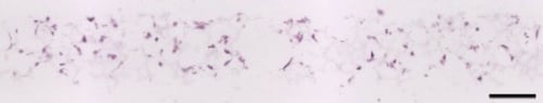
Figure 2. Brightfield micrograph showing the structure of CCD-18co cells cultured for 7 days on 15 mm diameter Alvetex Scaffold discs presented in 24 well plate format. Cells were fixed, embedded in paraffin wax, sectioned (10 µm) and counterstained with haematoxylin and eosin. Scale bar: 100 µm.
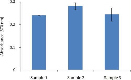
Figure 3. Biochemical analysis of cell viability using a standard MTT assay. Data from 3 sample replicates of CCD-18co cells are shown, each sampled in duplicate (n = 2, mean ± SD). Cells were cultured for 3 days on 15 mm Alvetex Scaffold discs presented in 24-well plate format.
3.2. 12 well plate format (AVP005)
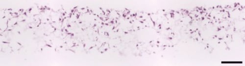
Figure 4. Brightfield micrograph showing the structure of CCD-18co cells cultured for 7 days on 15 mm diameter Alvetex Scaffold discs presented in 12 well insert in 12 well plate format. Cells were fixed, embedded in paraffin wax, sectioned (10 µm) and counterstained with haematoxylin and eosin. Scale bar: 100 µm.
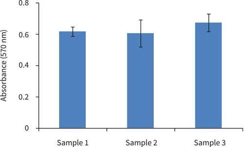
Figure 5. Biochemical analysis of cell viability using a standard MTT assay. Data from 3 sample replicates of CCD-18co cells are shown, each sampled in duplicates (n = 2, mean ± SD). Cells were cultured for 3 days on 15 mm Alvetex Scaffold discs presented in 12 well inserts in 12 well plate format.
4. References
- Grifin C et al, 2011. Pancratistatin selectively targets cancer cell mitochondria and reduces growth of human colon tumor xenografts. J Mol Cancer Ther 10(1):57-68.
- Takawa M et al, 2011 . Validation of the histone methyltransferase EZH2 as a therapeutic target for various types of human cancer and as a prognostic marker. Cancer Sci 102(7):1298- 305.
- Koshida Y et al, 2006. Interaction between stromal fibroblasts and colorectal cancer cells in the expression of vascular endothelial growth factor. J Surg Res 134(2):270-7.
- Koon HW et al, 2010. Substance P modulates colitis-associated fibrosis. Am J Pathol 177 (5):2300-9.
- Zhu Y et al, 2012. iNOS signaling interacts with COX-2 pathway in colonic fibroblasts. Exp Cell Res 318(16):2116-27.