Alvetex® Scaffold: Quick Start
● Download this protocol as a PDF (3 MB)
1. Alvetex Scaffold formats
Alvetex Scaffold is currently available in the following formats: 24 well plate (AVP006) and 12 well plate format (AVP002), suited to short-term experiments where full disc penetration is not required, 12 well inserts (AVP005) and 6 well inserts (AVP004), suitable for longer term and co-culture experiments and air/liquid interface set-ups. Inserts are compatible with standard tissue culture plates and with the Alvetex well insert holder in a deep Petri dish (AVP015), designed for demanding cell types to provide increased volumes of culture media. 12 well inserts fit into both 6 and 12 well plates (to fit into 12 well plates the extended arms must be snapped off). When deciding which Alvetex Scaffold format to use, the following factors should be considered in combination:
- Cell type and duration of experiment.
- The desired depth of cell penetration into the 3D scaffold.
- The type of assay or end point analysis to be performed.
2. Notes before starting
- Alvetex Scaffold is supplied sterile by gamma-irradiation and remains sterile until opened.
- Always handle Alvetex Scaffold with care, using gloves. Flat-ended forceps are recommended.
- Prior to use, Alvetex Scaffold must be rendered hydrophilic with a 70 % ethanol wash; add enough ethanol to completely cover the disc, remove, and wash 2× with appropriate culture media.
- To avoid drying, leave the disc in the final medium wash until required.
3. Seeding cells on Alvetex Scaffold
For optimal results, cells should be seeded directly onto the centre of the scaffold in a small volume of appropriate media to allow cell attachment. See Table 1 for specific details for each scaffold format.
In brief, when inoculating, aspirate off the wash medium thoroughly from the plate and carefully dispense cells on the middle of the disc. Replace the lid and incubate in a humidified incubator at 37 °C with 5 % CO2 for at least 30 minutes to facilitate cell attachment.
After this time, gently flood the wells with medium to the desired level, see Figure 2 and Table 2 for specific details for each scaffold format. With 3D cell culture there are likely to be many more cells growing per unit volume of medium, therefore media changes may be required more frequently than with 2D cultures.
4. Monitoring cell attachment and growth via light microscopy
Cell culture in Alvetex Scaffold allows the formation of multilayered, high-density cell populations which approximate the complexity and structure of in vivo tissues. When viewing an unstained, unsectioned Alvetex Scaffold 3D culture under a standard brightfield microscope, the combined density and thickness of the scaffold and the 3D culture within it prevent the clear visualisation of individual cells. However, to overcome this, common visible dyes such as Neutral Red staining solution can be successfully used to confirm cell attachment or to check confluency (Figure 1).
| Alvetex ® Scaffold format | Recommended cell seeding density* |
Recommended cell seeding volume |
Initial incubation period |
| 6 well insert (AVP004) in 6 well plate | 0.5–2.0 ×10⁶ | 100–150 µL | 30–90 minutes |
| 12 well insert (AVP005) in 6 well plate | 0.25–1.0 × 10⁶ | 50–75 µL | 30–90 minutes |
| 12 well insert (AVP005) in 12 well plate | 0.25–1.0 × 10⁶ | 50–75 µL | 30–90 minutes |
| 24 well insert (AVP012) in 12 well plate | 0.15–0.3 × 10⁶ | 50–75 µL | 30–180 minutes |
| 24 well insert (AVP012) in 24 well plate | 0.15–0.3 × 10⁶ | 50–75 µL | 30–180 minutes |
| 12 well plate format (AVP002) | 0.5–2.0 × 10⁶ | 100–150 µL | 30–180 minutes |
| 24 well plate format (AVP006) | 0.25–1.0 × 10⁶ | 50–75 µL | 30–180 minutes |
Table 1. Overview of the recommended cell seeding densities and volumes for the different Alvetex Scaffold formats.
* Optimal cell seeding density will be cell type specific.
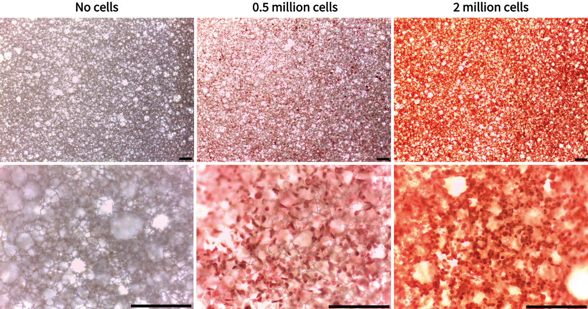
Figure 1. Microscopic appearance of CHO-K1 cells on Alvetex Scaffold after 24 hours as visualised by Neutral Red staining. Scale bar 200 µm. Note the increase in staining intensity with higher cell numbers.
|
 (ii.) Media from above and below |
(iii.) Media interconnected for routine 3D growth of cells with high metabolic activity/ proliferation rate. |
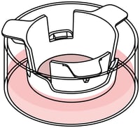
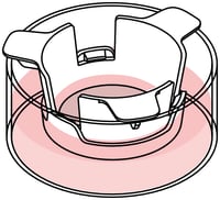
Figure 2. Media filling levels and well insert configurations.
| [A.] Using well insert (AVP004) or plate (AVP005) formats |
Media volumes for different feeding options | ||
| (i) Media from below only | (ii) Media from above and below | (iii) Media interconnected | |
| 6 well insert in 6 well plate | 3.5 ± 0.5 mL/well | 7 ± 1 mL/well | 10 ± 0.5 mL/well |
| 12 well insert in 6 well plate | 3.5 ± 0.5 mL/well | 7 ± 1 mL/well | 10 ± 0.5 mL/well |
| 12 well insert in 12 well plate | 1.6 ± 0.2 mL/well | 2.4 ± 0.2 mL/well | 3.8 ± 0.2 mL/well |
| [B.] Using 6 well insert (AVP004) with well insert holder (AVP015) | Feeding volume | ||
| (i) Media from below only | (ii) Media from above and below | (iii) Media interconnected | |
| 6 well insert in 6 well plate | 3.5 ± 0.5 mL/well | 7 ± 1 mL/well | 10 ± 0.5 mL/well |
| 12 well insert in 6 well plate | 3.5 ± 0.5 mL/well | 7 ± 1 mL/well | 10 ± 0.5 mL/well |
| 12 well insert in 12 well plate | 1.6 ± 0.2 mL/well | 2.4 ± 0.2 mL/well | 3.8 ± 0.2 mL/well |
| [C.] Using 12 well insert (AVP005) with well insert holder (AVP015) | Feeding volume | ||
| (i) Media from below only | (ii) Media from above and below | (iii) Media interconnected | |
| Low | 20 mL ± 1 mL | 40 mL ± 3 mL | 70 mL ± 3 mL |
| Medium | 34 mL ± 2 mL | 52 mL ± 3 mL | 82 mL ± 3 mL |
| High | 48 mL ± 2 mL | 70 mL ± 3 mL | 92 mL ± 3 mL |
Table 2 [A-C]. Feeding options for different Alvetex Scaffold formats.
5. Comparison of 3D cell growth patterns of HaCaT cells on Alvetex Scaffold presented in various formats
HaCaT cells were seeded (0.5 × 10⁶ cells per well) on Alvetex Scaffold in the following formats: 12 well plate (AVP002), 6 well inserts (AVP004) in 6 well plate and 12 well inserts (AVP005) in well insert holder in deep Petri dish (AVP015). Cultures were maintained for 7 days. After preserving in Bouin’s fixative the discs were paraffin embedded, sectioned (10 µm) and counterstained with haematoxylin and eosin.
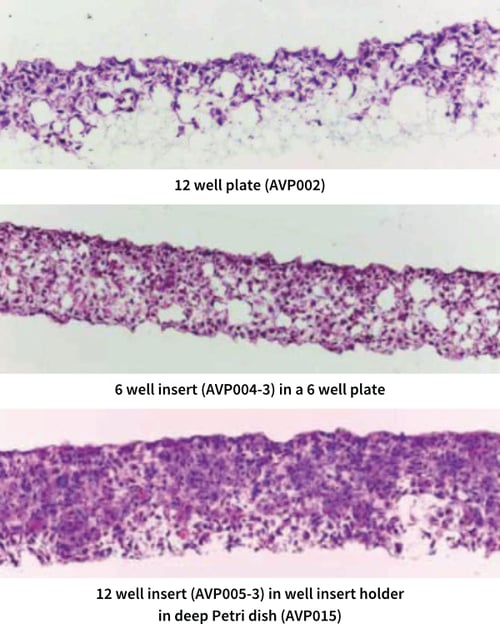
Figure 3. Comparison of 3D cell growth patterns of HaCaT cells cultured on various Alvetex Scaffold formats. Micrographs taken at 20× magnification.