Assay Description
REPROCELL has established three-dimensional (3D) autologous human skin equivalents (REPROSKIN) as physiologically relevant platforms for long-duration culture experiments in areas such as preclinical drug testing, cosmetic and skincare research, disease modelling, and precision medicine. REPROSKIN consists of human primary keratinocytes and fibroblasts, with options to include other cell types such as aged cells, immune cells or melanocytes.
We can engineer full thickness skin to investigate different aspects of cutaneous biology, as well as pathophysiology or drug-mediated responses in common skin disorders such as wounds, UV damage, atopic dermatitis, psoriasis and melanoma.
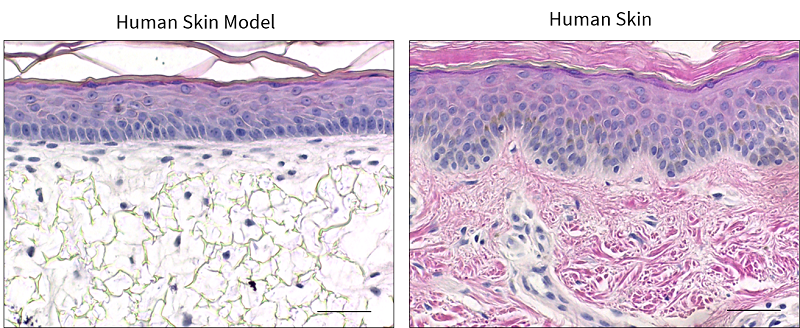 Figure 1. Histochemical analysis of our 3D human skin (REPROSKIN) compared with native human skin
Study Outline
3D skin models are developed through a coculture of primary keratinocytes and fibroblasts on Alvetex scaffolds in a serum-fortified medium at the air-liquid interface to optimize the self-assembly of human skin.
A description of the process to manufacture REPROSKIN is shown below. REPROCELL manufactures REPROSKIN as a custom order for immediate use in your project, with experiments conducted at REPROCELL's laboratories.

REPROSKIN is grown by a prolonged and natural 3D cell culture process that is required for biologically-accurate cell differentiation and expansion, as well as the expression of key epidermal differentiation markers, generation of dermo-epidermal junction (DEJ), and secretion of dermal-specific extracellular matrix (ECM) components. This natural biologically-driven process makes REPROSKIN the most human-relevant engineered skin on the market.
REPROSKIN can be used as a tool to address a wide range of biological questions in the presence of different test compounds or analytes to meet client requirements. On other hand, cell culture supernatants or conditioned media can be collected for various phenotypic analyses, such as multiplex ELISA (enzyme-linked immunosorbent assay) or liquid chromatography-mass spectrometry (LC/MS).
Experimental Design
An example of the experimental conditions for one test compound using REPROSKIN is presented below.
- Positive control
- Negative control (usually the test compound vehicle)
- Test compound(s) at various concentrations
- All above typically conducted in triplicate.
End Point Analysis
There are a range of endpoints that can be used to assess effectiveness in this system including:
- Barrier Integrity: Histological assessment, immunofluorescent staining, transepidermal water loss (TEWL) measurements
- Cytotoxicity: Histological assessment, TUNEL assay to detect and quantify apoptotic cells
- Biomarkers of Wound Repair: Histological assessment, immunofluorescent staining.
- Morphology of Wound Site: Gross Images, Histological assessment, immunofluorescent staining
- Pigmentary Changes: Melanin index, individual topography angle (ITA), Fontana-Masson histological staining
- Melanocyte Density: immunofluorescence detection and quantification of epidermal melanocytes
Testing information
Test compounds to be provided by the Sponsor in storable aliquots at the required test concentrations with information on the diluent/vehicle used. Stock solutions are typically prepared in deionized water unless otherwise requested. The sponsor also needs to provide sufficient test compound(s) to run the entire study.
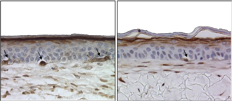 Figure 2: Histochemical analysis revealing migration of melanocytes (indicated by arrow) in our 3D skin model.


.jpg?width=756&height=425&name=Untitled%20design%20(10).jpg)