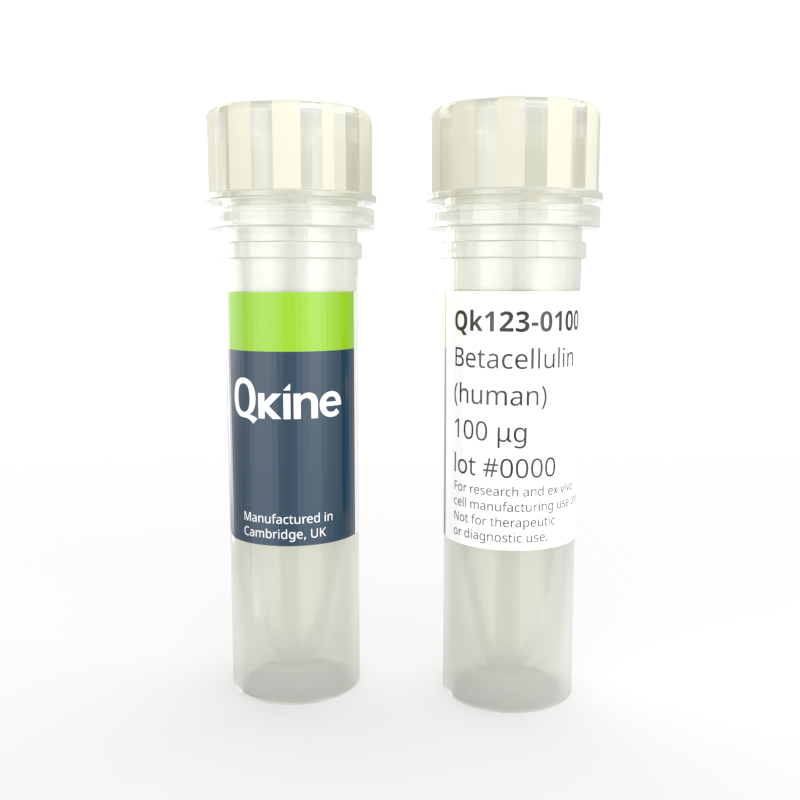
Recombinant human betacellulin
QK123
Brand: Qkine
Betacellulin is a member of the epidermal growth factor (EGF) family which regulates proliferation and differentiation in various stem cell types. Betacellulin stimulates proliferation and maintains pluripotency in neural and osteogenic stem cells. It can induce pancreatic β-cell differentiation in human embryonic stem cells (ESC) and differentiation of retinal progenitor cells.
Qkine human betacellulin is a highly pure and bioactive 9.89 kDa protein, animal origin-free (AOF), carrier-free, tag-free, and non-glycosylated to ensure its purity with exceptional lot-to-lot consistency.

Currency:
| Product name | Catalog number | Pack size | Price | Price (USD) | Price (GBP) | Price (EUR) |
|---|---|---|---|---|---|---|
| Recombinant human betacellulin, 25 µg | QK123-0025 | 25 µg | (select above) | $ 260.00 | £ 190.00 | € 222.00 |
| Recombinant human betacellulin, 50 µg | QK123-0050 | 50 µg | (select above) | $ 380.00 | £ 280.00 | € 328.00 |
| Recombinant human betacellulin, 100 µg | QK123-0100 | 100 µg | (select above) | $ 560.00 | £ 410.00 | € 479.00 |
| Recombinant human betacellulin, 500 µg | QK123-0500 | 500 µg | (select above) | $ 2225.00 | £ 1625.00 | € 1898.00 |
| Recombinant human betacellulin, 1000 µg | QK123-1000 | 1000 µg | (select above) | $ 3465.00 | £ 2530.00 | € 2956.00 |
Note: prices shown do not include shipping and handling charges.
Qkine company name and logo are the property of Qkine Ltd. UK.
Alternative protein names
Species reactivity
- human
- species similarity:
- mouse - 78%
- rat - 75%
- bovine - 90%
- porcine - 86%
Frequently used together:
Summary
- High purity human betacellulin (UniProt: P35070)
- 9.89 kDa
- >98%, by SDS-PAGE quantitative densitometry
- Expressed in E. coli
- Animal origin-free (AOF) and carrier protein-free
- Manufactured in our Cambridge, UK laboratories
- Lyophilized from acetonitrile
- Resuspend in 10 mM HCl (Reconstitution solution A) at >50 µg/ml, add carrier protein if desired, prepare single-use aliquots and store frozen at -20 °C (short-term) or -80 °C (long-term)
Featured applications
- Cellular proliferation, migration and survival
- Stimulation of angiogenesis and vascular network development
- Neurogenesis and neural differentiation
- Differentiation of retinal progenitor cells
Bioactivity
Recombinant betacellulin activity was determined using a luciferase assay. HEK293T cells were treated in triplicate with a serial dilution of betacellulin for 3 hours. Firefly activity was measured and normalized to the control Renilla luciferase activity. Data from Qk123 lot #204720. EC50 = 162.4 pg/ml.

Purity
Recombinant betacellulin migrates as a major band at approximately 15 kDa (monomer) in reduced (R) and non-reduced (NR) conditions. No contaminating protein bands are present. The purified recombinant protein 3 µg was resolved using 15% w/v SDS-PAGE in reduced (+β-mercaptoethanol, R) and non-reduced (NR) conditions and stained with Coomassie Brilliant Blue R250. Data from Qk123 lot #204740.

Further quality assays
- Mass spectrometry: single species with expected mass
- Recovery from stock vial: >95%
- Endotoxin: <0.005 EU/μg protein (below level of detection)
Protein background
Betacellulin was originally identified in conditioned media from mouse pancreatic-cell tumor cell line as a proliferation factor. It was discovered to be a member of the epidermal growth factor (EGF) family, binding the EGF receptor (EGF) [1, 2]. The BTC gene encodes a 178 amino acid precursor protein which is membrane anchored and cleaved to produce an 80 amino acid mature form. In addition to the EGFR, betacellulin is also a ligand for ERBB receptors, leading to activation of many intracellular signaling pathways including p38MAPK, PI3K/AKT, and JNK [1].
Initially identified as a pancreatic growth factor, betacellulin stimulates neogenesis and proliferation of pancreatic β-cells [1, 3], and pancreatic tumor cell lines [2]. It is able to covert selected non-β-cells into cells insulin producing cells, including mesenchymal stem cells (MSCs) [1, 5]. Betacellulin may also have a role in angiogenesis, it is detected in some models of retinal neovascularization and induces retinal vascular permeability and edema [1].
Betacellulin has been shown to have a regenerative role in the neural stem cell niche, stimulating proliferation and neurogenesis [5, 6]. In retinal progenitor cells (RPCs) betacellulin promotes proliferation and differentiation into retinal neurons [6]. However, in MSCs and pre-osteoblast cells it may increase proliferation but decrease osteogenic markers [7].
Background references
- Dahlhoff M, Wolf E and Schneider MR. The ABC of BTC: Structural properties and biological roles of betacellulin (2014), Seminars in Cell & Developmental Biology. https://doi.org/10.1016/j.semcdb.2014.01.002
- Kawaguchi M, Hosotani R, Kogire M, Ida J et al. Auto-induction and growth stimulatory effect of betacellulin in human pancreatic cancer cells (2000), International Journal of Oncology. PMID: 10601546
- Yamamoto K, Miyagawa J, M Waguri M et al. Recombinant human betacellulin promotes the neogenesis of beta-cells and ameliorates glucose intolerance in mice with diabetes induced by selective alloxan perfusion (2000), Diabetes. https://doi.org/10.2337/diabetes.49.12.2021
- Paz AH, Salton GD, Ayala-Lugo A et al. Betacellulin Overexpression in Mesenchymal Stem Cells Induces Insulin Secretion In Vitro and Ameliorates Streptozotocin-Induced Hyperglycemia in Rats (2010), Stem Cells and Development. https://doi.org/10.1089/scd.2009.0490
- Gómez-Gaviro MV, Scott CE, Sesay AK et al. Betacellulin promotes cell proliferation in the neural stem cell niche and stimulates neurogenesis (2012), Proc. Natl. Acad. Sci. U.S.A. https://doi.org/10.1073/pnas.1016199109
- Zhang D, Shen B, Zhang Y et al. Betacellulin regulates the proliferation and differentiation of retinal progenitor cells in vitro (2017) Journal of Cellular and Molecular Medicine. https://doi.org/10.1111/jcmm.13321
- Genetos DC, Rao RR and Vidal MA. Betacellulin inhibits osteogenic differentiation and stimulates proliferation through HIF-1α (2010), Cell Tissue Res. https://doi.org/10.1007/s00441-010-0929-0