Histology (1): Choosing the Right Fixative to Preserve 3D Cell Cultures
● Download this protocol as a PDF (7.4 MB)
1. Introduction
These histology protocols contain a series of detailed methods that will allow the user to examine the morphology of their cultured cells subsequent to 3D growth. These are not in any way exhaustive and there is scope to include additional methods as the user deems appropriate. Users should also perform their own risk assessments as safety evaluations contained in these protocols are for information only.
2. Fixation methods
Fixation is performed in order to preserve the tissue or culture from the putrefaction (destruction by micro-organisms), autolysis (self-digestion by lysosomal enzymes) and abnormal metabolism of the isolated tissues deprived of oxygen and nutrients. Fixation of 3D cultures in Alvetex Scaffold can be achieved by chemical fixation. The use of uncontrolled high heat by flame is not recommended.
Fixation is usually the first stage in a multistep process to prepare a sample for microscopy or other analysis. So, when choosing a fixative, the final purpose of the section must be considered and matched by the requirements for the analytical technique. Some fixatives are suitable for general structure analysis, others for evaluating cytological detail and some for histochemistry. Survival of tissue antigens for immunochemical staining depends on the type and concentration of fixative, on fixation time, and on the size of the tissue specimen to be fixed. Given the thin nature of Alvetex Scaffold, fixation of cells is rapid, uniform and efficient, preserving the 3D culture in a life-like condition.
Recommended chemical fixatives for tissue structures grown in Alvetex Scaffold are:
2.1. Bouin’s Fixative
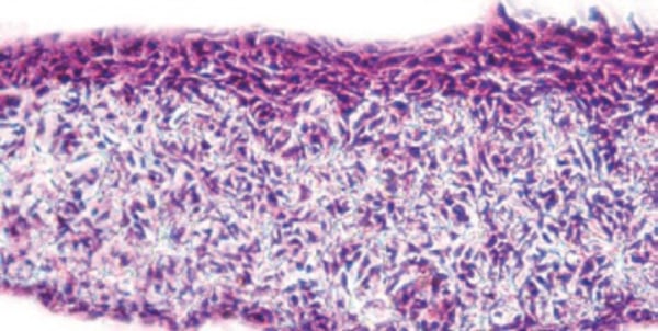
Is an excellent fixative for use when samples are to be paraffin embedded, sectioned and stained for general histology (see Figure 1.), especially for the trichrome stains. Bouin’s fixation is not recommended for immuno-detection. Do not use Bouin’s fixative before in situ hybridisation as it will prevent RNA from being detected.
2.1.1. Method
- Working in a fume cupboard wearing appropriate Personal Protective Equipment (PPE) measure out saturated picric acid (750 mL), formaldehyde (250 mL) and glacial acetic acid (50 mL).
- Mix well in a 1 litre Duran bottle. Fixative is stable for 1 year at room temperature.
2.1.2. Safety precautions
CARCINOGENIC, IRRITANT, CORROSIVE and TOXIC
2.2. Paraformaldehyde (4 % PFA)
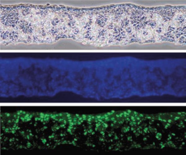
Suitable for paraffin embedding and sectioning. It is the fixative of choice for immunocytochemical analysis. Samples can also be stained for general histology but the degree of fixation is less vigorous than Bouin’s so the quality of the morphology obtained will be less. This fixative allows for subsequent immuno-detection of certain antigens and should therefore be used when the objective is to study morphology and protein expression simultaneously. (See Figure 2.)
2.1.1. Method
- Working in a fume cupboard wearing appropriate PPE heat up 1 litre of Phosphate Buffered Saline (PBS) to 60 °C on a hotplate stirrer.
- Add 40 g of paraformaldehyde (PFA) powder to the warm PBS. Use a magnetic stirrer and hotplate to dissolve PFA.
- Slowly add drops of 1 M sodium hydroxide solution until the solution is clear.
- Filter the fixative and adjust pH to 7.3-7.4.
- Allow fixative to cool to 5 ± 3 °C before use.
- Aliquots can be stored at –20 °C with a shelf life of 6 months.
2.1.2. Safety precautions
CARCINOGENIC, IRRITANT, CORROSIVE and TOXIC
2.3. Karnovsky’s fixative
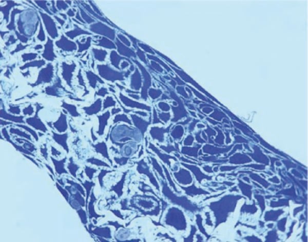
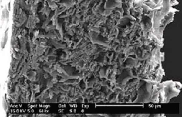
Karnovsky’s fixative is a mixture of paraformaldehyde and glutaraldehyde. It is suitable for use when preparing samples for light microscopy in resin embedding and sectioning, and for electron microscopy. This fixative should always be prepared fresh. (See Figures 3 and 4 respectively).
2.3.1. Method
Karnovsky’s fixative is 2 % paraformaldehyde, 2.5 % glutaraldehyde in 0.1 M phosphate buffer pH 7.4.
- Prepare 8 % PFA:
- Working in a fume cupboard wearing appropriate PPE heat up 90 mL of water to 65°C on a hotplate stirrer.
- Add 8 g of paraformaldehyde (PFA) powder to the warm water. Use a magnetic stirrer and hotplate to dissolve PFA (approximately 15-20 minutes).
- Slowly add drops of 1 M sodium hydroxide solution until the solution is clear. Make up to 100 mL with additional water.
- Filter the fixative and adjust pH to 7.3-7.4.
- Allow fixative to cool to room temperature. Store at –20 °C in aliquots. Frozen aliquots are stable for up to 6 months.
- Dispense 25 mL for use in fixative.
- Prepare 0.2 M phosphate buffer:
- Weigh out 35.61 g of disodium monohydrogen phosphate (Na2HPO4.2H2O) – make up to 1000 mL with distilled H2O, stir until dissolved (Solution X). 1 year shelf life at room temperature.
- Weigh out 27.6 g of sodium dihydrogen phosphate (NaH2PO4.H2O) – make up to 1000 mL with distilled H2O, stir until dissolved (Solution Y). 1 year shelf life at room temperature.
- Add 40.5 mL of Solution X to 9.5 mL of Solution Y to give 50 mL 0.2M phosphate buffer and adjust to pH 7.4. This is to be used fresh.
- Prepare Karnovsky’s Fixative (for 100 ml add the following together):
- Mix together 8 % paraformaldehyde (25 mL), 25 % glutaraldehyde (Fluka 49362 or equivalent, 10 mL) and 0.2 M phosphate buffer (50 mL). Make up to 100 mL with distilled water.
- This is to be used fresh.
2.3.2. Safety precautions
CARCINOGENIC, IRRITANT, CORROSIVE and TOXIC
2.4. Buffered osmium tetroxide fixative
Buffered osmium tetroxide fixative is suitable as a second-stage fixative when preparing samples for electron microscopy. It is also used when samples are to be resin embedded and sectioned. This fixative increases the samples bulk conductivity, which is required for scanning electron microscopy. (See Figures 3 and 4 respectively).
2.4.1. Method
- Working in a fume hood throughout and wearing appropriate PPE, combine equal amounts of the 2 % osmium tetroxide solution and 0.2 M phosphate buffer (for recipe see above) in a clean 15 mL centrifuge tube.
- Vortex gently.
- Store in a fume hood at room temperature.
- Final concentrations: 1 % osmium tetroxide in 0.1 M phosphate buffer. Cacodylate buffer may be used to replace the phosphate buffer.
2.4.2. Safety precautions
CARCINOGENIC, IRRITANT, CORROSIVE and TOXIC
Note: Osmium tetroxide is highly toxic and volatile and should be used and stored only in a chemical fume hood.
3. Examples
Below are examples of how the same 3D culture of human keratinocytes (HaCaT) can be processed and subsequently imaged in alternative ways:

Figure 1. Human keratinocyte cell line (HaCaT) grown in Alvetex Scaffold (7 days air exposure). The subsequent culture was fixed in Bouin’s fixative and processed for paraffin wax embedding and sectioning. The sectioned materials (10 µm) were stained with Haematoxylin and Eosin for morphological analysis.

Figure 2. Human keratinocyte cell line (HaCaT) grown in Alvetex Scaffold (7 days air
exposure). The subsequent culture was fixed in 4 % paraformaldehyde and processed for paraffin wax embedding and immunohistochemical analysis by fluorescent microscopy. The three images from the same region show; phase (top), blue fluorescent Hoescht 33258 nuclei stain (middle) and Ki67 staining (bottom).

Figure 3. Human keratinocyte cell line (HaCaT) grown in Alvetex Scaffold (7 days air exposure). The subsequent culture was primarily fixed in Karnovsky’s fixative followed by a secondary fixative osmium tetroxide and processed for resin embedding (L R White resin). Resin sections (1 µm) were stained with Toluidene Blue for structural analysis by light microscopy.

Figure 4. Human keratinocyte cell line (HaCaT) grown in Alvetex Scaffold (28 days air exposure). The subsequent culture was primarily fixed in Karnovsky’s fixative followed by a secondary fixative osmium tetroxide and processed for structural analysis by scanning electron microscopy. For scale see bar inserts.