Neuronal Differentiation Services
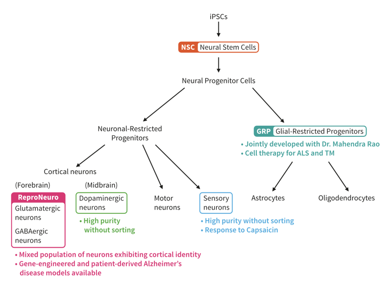
Patient-specific iPSCs are a self-renewing and expandable cell population with the potential to differentiate into virtually any cell type. The ability to model human diseases using iPSCs has revolutionized the ways in which we study monogenic, complex, and epigenetic disorders, as well as early- and late-onset diseases.
ReproNeuro Technology
REPROCELL was one of the first companies to make differentiated cell types commercially available. We launched our first commercial neurons, ReproNeuro, in 2012, and are now in possession of a wide range of iPSC differentiation capabilities. Currently, REPROCELL is using ReproNeuro technology to expand our range of capabilities even further — our most recent development being the introduction of dopaminergic and sensory neurons.
With ReproNeuro, you can access:
✓ Neurons derived from human iPSCs
✓ Differentiated cells that express neuron-specific markers, including drebrin
✓ Neurons that are functional with measurable outputs
✓ iPSCs for modelling Alzheimer’s disease
✓ iPSC-derived sensory neurons and dopaminergic neurons
Discover Our Sensory Neurons

Expression of Neuron-Specific Markers in ReproNeuro
ReproNeuro is a mixed population of forebrain and midbrain neurons that express various neuron-specific markers (Figure 1). This feature makes ReproNeuro the ideal model for investigating neurological systems.
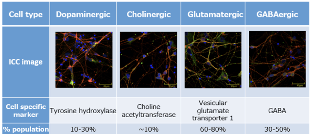
Figure 1: ReproNeuro contains a mix of neuron types and therefore mimics in vivo conditions.
In this study immunostaining was used to determine the percentage population of each neuron type. BIII tubulin was stained red, nuclei stained blue, and cell specific markers were tagged with a green fluorophore.
Differentiated Cells Express Drebin, the Neuron-Specific Marker
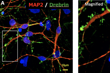
Drebrin, a marker of synapse formation related to neuroplasticity, is expressed by ReproNeuro cells. This protein, which marks the formation of synapses and neuritic spines, is co-localized with the presynaptic protein Vglut2 on ReproNeuro cells (Figure 2).
 |
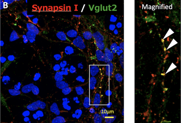 |
Figure 2: ReproNeuro was evaluated for the expression of Drebrin via immunostaining.
(A.) After two weeks, a spine-like accumulation of drebrin (dreen) was observed on the neuritic protrusions of ReproNeuro cells. (B.) This was found to be co-localised with the presynaptic protein Vglut2, kindly provide by Dr Shirao, Gunma University.
ReproNeuro Cells Function Like Neurons
After 14 days culture, our scientists added Fluo-8 to ReproNeuro cells with the aim of observing changes in calcium (Ca2+) concentration. Almost all cells in the culture displayed spontaneous neural activity in response to glutamic acid — illustrated beautifully in this video by Keio University (figure 3).
Figure 3: Calcium indicator Fluo-8 demonstrates spontaneous electrical activity of ReproNeuro cells.
Video curtesy of Prof. Oka and Mr. Enya in Keio University
When cultured in ReproNeuro MQ Medium, differentiated cells also respond to electrical stimulation (Figure 4), glutamic acid (Figure 5A), and show evidence of functional sodium (Na+) channels (Figure 5B). The use of Neuro MQ Medium meant that these iPSC cells matured quicker, and therefore displayed functional properties as early as two weeks post-differentiation.
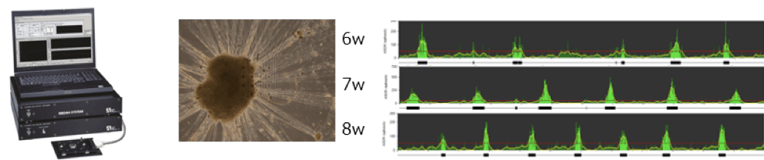
Figure 4: Multielectrode Array (MEA) Assay.
ReproNeuro cells were cultured in ReproNeuro MQ Medium on electrode plates and signals due to neural activity were obtained. Mature functional phenotype can be observed as early as week two, with the signal intensifying after six weeks culture.
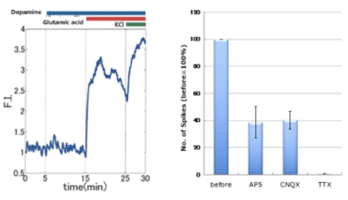
Figure 5: ReproNeuro cells respond to glutamic acid and possess functional sodium channels.
(A.) Our iPSC-derived neurons respond to glutamic acid. (B.) Assessment of the inhibition effects of various glutamate receptor antagonists (APS, CNQX, TTX) demonstrating Na+ channel activity.
Customized Disease Modelling
Thanks to our partner company GenAhead Bio®, it is now possible for REPROCELL to genetically modify your iPSCs using advanced CRISPR-SNIPER gene-editing technology. We can now generate patient-derived iPSCs that possess your desired mutation, or repair DNA mutations for the purpose of transplantation.
ReproNeuro AD: Neurons for Modelling Alzheimer’s Disease
At REPROCELL we provide two functional neuronal models for Alzheimer’s Disease (AD): ReproNeuro AD-mutation, and ReproNeuro AD-patient 1. Our first model contains neurons differentiated from iPSCs transfected with the P117L mutant variant of the AD gene Presenilin 1 (PS1). In contrast, our ReproNeuro AD-patient 1 was developed from patient-derived iPSCs donated by a carrier of the R62H mutant variant of the Presenilin 2 (PS2) gene. This means we can offer both genetically modified and patient-derived models of AD.
REPROCELL CASE STUDY
Modelling Alzheimer’s Disease with ReproNeuro
In this study, our scientists aimed to determine whether ReproNeuro AD-patient 1 and ReproNeuro AD-mutation displayed the phenotypical characteristics of Alzheimer’s disease (AD) compared with our healthy ReproNeuro cells. To achieve this, the neuritic protrusions present on all three cells types were assessed via immunocytochemistry (Figure 6A). These results were subsequently quantified so that they could be compared (Figure 6B). Our scientists found that these characteristic structures were present in both AD models, as observed below.
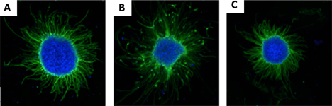
Figure 6: Formation and elongation of neuritic protrusions in three different cell models. (A.) ReproNeuro (B.) Patient-derived AD model (C.) Gene-modified AD model.
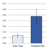
Generation of amyloid beta peptides
In our final experiment, the concentration of amyloid beta (Aβ) contained in the extracellular fluid of ReproNeuro AD-mutation cells was assessed. This confirmed that the ratio of Aβ-42 generated was higher in REPROCELL’s model system compared with wild-type neurons (right figure). This model system would therefore be superior in studies to wild-type PS1 mutated neurons in testing the amyloid hypothesis.
Custom Generation of Sensory and Dopaminergic Neurons
Custom generation of other neuron types is available upon request. Below, you can find an example case study of custom iPSCs generated in partnership with one of our clients, FANCL. Our differentiation technique provides high purity neurons which can be used to model a range of nervous conditions — from bipolar disorder to Parkinson’s disease (Figure 7).
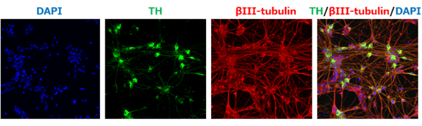
Figure 7: For the study of Parkinson’s disease, we offer iPSC-derived dopaminergic (DA) neurons. Using our ReproNeuro-derived dopaminergicneurons were generated at high purity without sorting. Immunostaining was used to identify tyrosine hydroxylase (TH)-positive (green) cells. As TH is a DA neuron marker, these results indicated that the differentiation process was up to 60% efficient without sorting.
REPROCELL CASE STUDY
Differentiation of Sensory Neurons
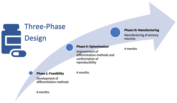
In 2019, FANCL asked REPROCELL to develop sensory neurons derived from human iPSCs. The project had a three-step design, beginning with a pilot study to test feasibility and ending with product manufacturing.
These studies led to the production of neurocytes that expressed both peripheral and sensory neuron markers (Figure 5) which responded to 100 nm capsaicin – indicating the presence of a sensory response to nociceptive stimuli.
A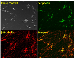
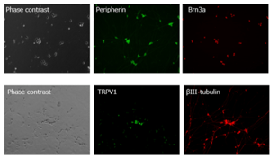
Figure 8: Immunostaining experiments revealed that the resultant neurocytes express PNS and sensory neuron markers.
(A.) Almost all cells express the PNS marker (green) peripherin and (B.) sensory neuron markers Brn3a (red) and TRPV1 (green). These neurons were developed in partnership with FANCL Co., Ltd.


