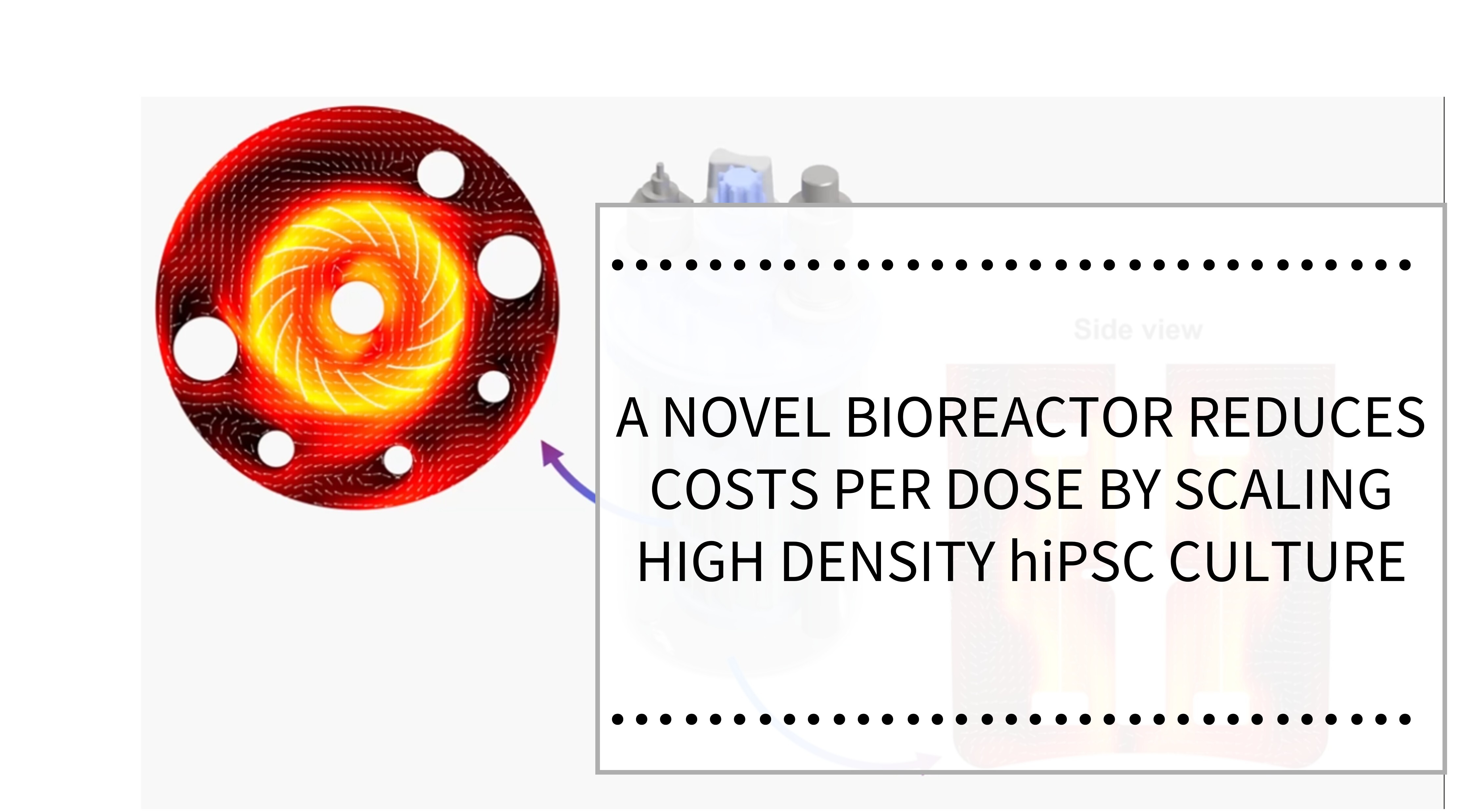
Induced Pluripotent Stem Cells (iPSCs) are revolutionizing regenerative medicine. They play a pivotal role in disease modeling and modern cell therapy development. One of the challenges with cell-based models is scale: How do we get sufficient cells at an affordable cost? Suspension cultures using bioreactors offer a solution.
Bioreactors are available from several vendors in scales from 5 mL to more than 2000 L. Smaller, single-use, lab-scale bioreactors of up to 500 mL are ideal for optimizing the conditions of cell growth and producing 106 to 109 cells in a single bioreactor, using a cost-effective amount of media.
A major supplier of single-use lab-scale bioreactors is Able Biott (Tokyo, Japan). Their bioreactors are available from REPROCELL (https://www.reprocell.com/product-catalog/able-biott-bioreactor-systems) in 5 mL to 500 mL sizes. These bioreactors have a proprietary impeller design, optimized for iPSC culture, and are designed to go into a standard cell incubator to provide temperature, atmosphere, and humidity control.
Video 1. ABLE Biott 3D Magnetic Stir and Disposable Bioreactor System
Able Biott bioreactors have been used in several ways to support regenerative medicine and disease model development. Examples include cardiomyocyte sheets to treat ischemic cardiomyopathy, neural organoids as a model for Alzheimer’s disease, and scale-up optimization for creating cell banks of iPSCs.
Cardiomyocyte Sheets
The use of iPSCs to develop cardiomyocyte sheets (CSs) for the treatment of ischemic cardiomyopathy (“heart attack”) has been a subject of intense research since before 2012[1]. Several recent studies have demonstrated the advantage of using Able Biott bioreactors in the early differentiation protocol to improve the yield and quality of the cardiomyocyte sheets.
A recent study[2] from Osaka University examined the effects of the synthetic prostacyclin, ONO-1301, which has been shown to promote angiogenesis, on the development of functional CS. iPSCs were initially cultured in Primate ES Medium (REPROCELL Cat No RCHEMD001) and expanded and differentiated into cardiomyocytes in Able Biott bioreactors. The cardiomyocytes were transferred to scaffolds with or without a slow-release form of ONO-1301 and allowed to develop.
The cells were then transplanted into a rat model of myocardial infarction (MI). Both treated (with ONO-1301) and control groups showed microscopic and immunoassay indicators of CS development. In addition, the treated group demonstrated increased proangiogenic signal expression, and the implanted treated CS group showed greater survival and improved recovery of cardiac ejection fraction in vivo (Figure 1). This study demonstrated that the ONO-1301-based protocol generates enhanced angiogenesis, survival of CS, and improved cardiac function.

Figure 1. Treatment with ONO-1301 caused improved ejection fraction in a rat model of myocardial infarction. Green: ONO-treated; Blue: ONO negative control; Grey: Sham control. Cardiomyocytes were differentiated in an Able Biott bioreactor prior to implantation in a nude rat model.
An earlier study from Nippon Medical School used Able Biott bioreactors to expand embryoid bodies (EBs) and differentiate them into cardiomyocytes created from genetically modified iPSCs[3]. CSs were developed on treated wells of a plate and characterized microscopically and by electrophysiology assays (Figure 2). This method produced CS that had functional ion channels, gap junctions, and mature sarcomeres after more than 150 days of incubation.

Figure 2. Gene-edited iPSC-derived cardiomyocytes were developed in bioreactors, transferred to 2D culture to develop CS over 79 days in culture. Different color curves represent separate biological replicates. The CS were functionally tested by measuring beating activity by Ca2+ uptake, showing synchronous beating.
Brain Organoids
Human cellular models of Alzheimer’s disease (AD) and tauopathies are generally limited to replicating only the earliest stages of disease progression. To address these limitations, scientists at Keio University School of Medicine in Tokyo developed a method to generate forebrain organoids (FBOs) from iPSCs derived from familial AD (fAD) patients and healthy controls by modulating fibroblast growth factor 2 (FGF2) concentrations[4]. This method uses the Able Biott bioreactors to scale up EB intermediates and differentiate them into FBOs.
These fAD-derived FBOs recapitulated key features of early AD pathology, including amyloid-β accumulation (Figure 3) and increased tau phosphorylation, although they did not develop tau aggregates. To model full tau pathology, adenovirus expressing the P301L mutant tau was introduced into the FBOs. In these Tau-P301L FBOs, hallmark features of tauopathy, including tau fibrils in neuronal cell bodies and neurites as visualized by immunoelectron microscopy, were observed.This advanced FBO model offers a robust platform for studying the pathogenic mechanisms underlying tauopathy and evaluating potential therapeutic targets in drug discovery.

Figure 3. The ratio of Aβ42 to Aβ40 in the culture medium of 120-day FBOs, as measured using ELISA. Error bars represent ± SD. This result shows that fAD FBOs express higher levels of Aβ42 compared to control FBOs.
iPSC Scaleup
The clinical applicability of human iPSCs depends on a range of critical quality attributes. These include the development of high-quality cell banks, long-term genomic stability, and the maintenance of pluripotency. In a new study[5], the cellular and genomic stability of iPSC lines, that were generated and cryopreserved under cGMP conditions five years prior, were reassessed. Able Biott bioreactors were used for the post-thaw expansion of the cells, and the iPSC lines were evaluated for pluripotency and differentiated into representative cell types of all three germ layers: cardiomyocytes (CMs), neural stem cells (NSCs), and definitive endoderm (DE). To further assess their expansion potential, cells were cultured under both two-dimensional (2D) and three-dimensional (3D) conditions.
All three iPSC lines thawed efficiently, adhered to L7™ matrix, and formed characteristic iPSC colonies expressing key pluripotency markers for at least 15 passages. The lines retained the ability to undergo both spontaneous and directed differentiation into the three germ layers, as demonstrated by the expression of lineage-specific markers. Post-thaw assessments confirmed normal karyotypes and negative results for mycoplasma contamination and sterility testing. Crucially, the cells maintained robust proliferative capacity in both 2D and 3D cultures without evidence of genomic instability, loss of pluripotency, or diminished telomerase activity. These findings highlight the long-term stability, functionality, and clinical relevance of cGMP-compliant iPSC lines, supporting their viability as a dependable and scalable source of starting material to produce iPSC-derived cell therapies. In addition, they highlight the utility of Able Biott bioreactors in evaluating the suitability of iPSCs for GMP manufacturing.
Conclusion
These three examples highlight the utility of lab scale bioreactors such as Able Biott in cell therapy development. Information about all of our Able Biott products can be found at https://www.reprocell.com/product-catalog/able-biott-bioreactor-systems or via email info-us@reprocell.com.
References
1. Kawamura M, et al. “Feasibility, safety, and therapeutic efficacy of human induced pluripotent stem cell-derived cardiomyocyte sheets in a porcine ischemic cardiomyopathy model.” Circulation 126:S29 (2012) https://doi.org/10.1161/CIRCULATIONAHA.111.084343
2. Qu X, et al., “ONO-1301 enhances post-transplantation survival of human induced pluripotent stem cell-derived cardiac tissue sheet by promoting angiogenesis.” J Heart Lung Transplant 42:716 (2023) https://doi.org/10.1016/j.healun.2023.01.018
3. Aoyama J, et al. “Spatiotemporal imaging documented the maturation of the cardiomyocytes from human induced pluripotent stem cells.” J Thoracic Cardiovasc Surg 159:2260 (2020) https://doi.org/10.1016/j.jtcvs.2019.06.060
4. Shimada H et al. “A next-generation iPSC-derived forebrain organoid model of tauopathy with tau fibrils by AAV-mediated gene transfer.” Cell Rep Methods 2:100289 (2022). 10.1016/j.crmeth.2022.100289
5. Shafa M, et al. “Long-term stability and differentiation potential of cryopreserved cGMP-compliant human induced pluripotent stem cells.” Int J Mol Sci 21:108 (2020). https://doi.org/10.3390/ijms21010108
About the author
Robert Annand PhD, Senior Technical Product Manager, REPROCELL USA
Before the REPROCELL merger, Robert was the Technical Product Manager at Stemgent. He dreams of retiring with his wife to many years of happiness and health, after helping make the world a better place. You can contact Robert on LinkedIn.
Subjects we write about
- 3D Cell Culture
- Cell Culture
- Central Lab Services
- Clinical Capabilities
- Disease Modeling
- Drug Discovery
- Gene Editing
- Genomic Services
- GMP
- Human Tissue Samples
- Human Tissue Testing
- IBD
- Life Sciences
- Master Cell Banks
- Neurons
- Oligonucleotide Synthesis
- Pharmacology-AI
- Precision Medicine
- Product Catalog
- Regenerative Medicine
- Respiratory Disease
- Safety Pharmacology
- Skin Disease
- Stem Cells


.png?width=400&height=225&name=hubspot_featured_image_1200x628%20(1).png)