Example Protocol for the Culture of the MCF-10A Cell Line on Alvetex® Scaffold 12 Well Insert Format and 96 Well Plate Format
● Download this protocol as a PDF (3.5 MB)
1. Introduction
Alvetex Scaffold is available in several cell culture formats including 24 well plate (AVP006), 12 well plate (AVP002), 6 well insert (AVP004), 12 well insert (AVP005), and 24 well insert (AVP012).
24 well and 12 well plates are suitable for shorter term cultures and for applications where limited cell penetration into the scaffold is required. Well insert formats generally support longer term cultures and deeper cell penetration into the scaffold. They also provide for conveniently tailored media set ups (see the Alvetex Scaffold Quick Start protocol).
The availability of two different well insert formats enables choice on the basis of desired culture size and cell expenditure. 6 well inserts can be placed in conventional 6 well plates, while 12 well inserts can be placed in either 6 well plates or 12 well plates, depending on media requirements. Alternatively, both insert types can be housed in the dedicated Well Insert Holder in Deep Well Petri Dish (AVP015) to allow for increased media volumes and prolonged cell culture.
The MCF-10A cell line was derived the breast tissue of a Caucasian woman with fibrocystic disease. The cell line is non-tumorigenic, and is used as a model to study breast glandular epithelial development and neoplastic transformation[1,2].
2. Methods
2.1. Preparation for 3D cell culture in Alvetex Scaffold
- MCF-10A cells (ATCC, CRL-10317) were routinely maintained in T-75 flasks.
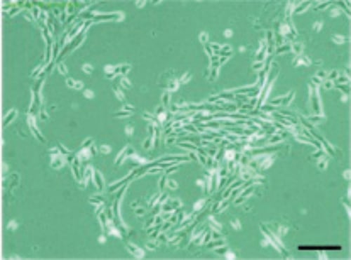
Figure 1. Phase contrast micrograph of MCF-10A cells grown in conventional 2D culture plates.
Scale bar: 200 µm.
-
Complete media consisted of: MEBMTM basal medium (Lonza, CC-3151) supplemented with the MEGM™ Single Quot Kit (Lonza, CC-4136).
-
Cells were harvested by trypsinisation according to the supplier’s instructions and counted using Trypan Blue exclusion.
-
Cells were resuspended in complete culture medium according to desired seeding densities (see specific examples below).
-
Consult example protocols below for culture of MCF-10A cells in either 12 well insert (AVP005) or 96 well plate (AVP009) formats.
2.2. 12 well Insert Format (AVP005)
-
Alvetex Scaffold 12 well inserts were prepared for seeding by dipping once in 70 % ethanol and twice in complete culture medium. Prepared inserts were placed into standard 6 well plates.
-
1 × 10⁶ cells in 75 µL of complete culture medium were added to the centre of each Alvetex Scaffold disc.
-
Each plate was incubated for 30 minutes at 37 °C with 5 % CO2 to allow cells to settle into the scaffold.
-
10 mL of complete culture medium was carefully added to each well, taking care not to dislodge cells from the Alvetex Scaffold.
-
Plates were re-incubated and maintained by complete media exchange every 2-3 days.
Note: This method can be applied to the use of Alvetex Scaffold in 6 well insert format (AVP004). Adjust cell seeding and media volumes according to the guidelines provided in the Quick Start Protocol.
2.3. 96 well Plate Format (AVP009)
-
Alvetex Scaffold 96 well plates were prepared for cell seeding with a 70 % ethanol wash (50 µL per well) and two washes with PBS (100 µL per well). Complete culture medium was added to each well (100 µL) and plates were incubated at 37 °C with 5 % CO2 until cells were ready for seeding.
-
Prior to cell seeding, media was aspirated from each well.
-
Cells were introduced in a total volume of 200 µL per well, at a range of seeding densities from 1 × 10⁴ to 1 × 10⁵ cells per well.
-
Plates were incubated at 37 °C with 5 % CO2 and maintained by complete media exchange every day, for up to 7 days total.
Note: This method can be applied to the use of Alvetex Scaffold in either 12 well plate (AVP002) or 24 well plate (AVP006) formats by adjusting cell seeding and media volumes accordingly.
3. Example Data
3.1. 12 well Insert Format (AVP005)
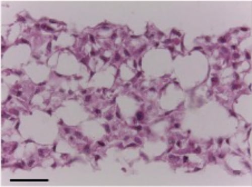
Figure 2. Brightfield micrographs showing the structure of MCF-10A cells cultured for 7 days on 15 mm diameter. Alvetex Scaffold discs in 12 well inserts placed in 6-well plates. Cells were fixed, embedded in paraffin wax, sectioned (10 µm) and counterstained with haematoxylin and eosin. Scale bar: 50 µm.
3.2. 96 well Plate Format (AVP009)
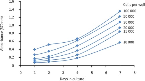
Figure 3. MTT cell viability curves tracking the progress of MCF-10A cultures seeded at different densities in Alvetex Scaffold 96-well plate format.
4. References
- Debnath J, et al. Morphogenesis and oncogenesis of MCF-10A mammary epithelial acini grown in three dimensional basement membrane cultures. Methods 2003. 30: 256-68.
- Pauley RJ, et al. The MCF-10 family of spontaneously immortalized human breast epithelial cell lines: models of neoplastic progression. Eur J Cancer Prev 1993. 3: 67-76.