Precision-Cut Lung Slice (PCLS) Culture
Drug Discovery Assay – reference number: B112
| Assay type: | Respiratory |
| Tissue: | Human precision-cut lung slice (PCLS) |
| Target: | Glucocorticoid receptors/ retinoid receptors/ PDE4 |
| Control compound: | Fluticasone/ roflumilast |
| Study type: | Ex vivo cultures |
| Functional endpoint: | Cytokine release |
Testing Information
Introduction
This experiment assesses whether test articles cause a reduction in cytokine release from precision-cut lung slices (PCLS). The specific results that will be provided are the effects of test articles on the levels of TNF-alpha release in human PCLS.
Test Article Requirements
Test article(s) to be provided by the Sponsor in storable aliquots at required test concentrations with information on diluent vehicle used. Stock solutions are prepared in deionized water unless otherwise requested. Sponsor to provide sufficient test article to run the entire study.
Suggested Testing
A minimum of triplicate per condition. A high number of slices are available per donor.
Study Outline: Rationale and Experimental Design
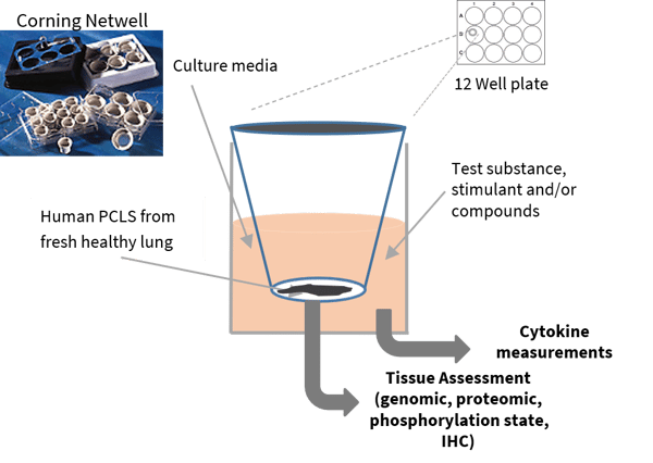
Core biopsies are created from healthy or diseased lung tissue samples stabilized in agarose. PCLS are created at a thickness of 250 or 500 µm and placed on a mesh in fortified media before being submerged in a 12 well plate in optimum conditions. Slices remain viable in culture for up to 7 days as shown by histology (Masson’s trichrome) and MTT turnover.
An example of the conditions assessed for 3 test articles are detailed below (it is recommended a minimum of triplicates should be used for each condition):
- Non-stimulated control
- Stimulated control
- Test article 1
- Test article 2
- Test article 3
- Positive control

Exclusion Criteria
No specific exclusion criteria are in place other than to reject macroscopically diseased/necrotic tissue.
Standardization and Qualification
Tissue that does not respond to stimulation with LPS would be deemed non-functional.
Analysis
Cytokine analysis: Supernatant samples can be analyzed for specific analytes of your choice by a multiplex ELISA platform. Each analyte will be quantified by interpolation against a standard curve generated on the same 96 well analysis plate.
Other forms of end point analysis are available such as gene expression or immunohistochemistry.
Example data from PCLS culture studies
Viable tissue as supported by Isoprenaline-induced bronchodilation

Tissue biopsies and slices can be maintained in culture for hours to days, allowing a wide range of functional endpoints to be measured. A series of images can be captured by video microscopy and image analysis allowing measurements of lumen diameter; here, a time-capture series of human isolated bronchus demonstrates relaxation upon exposure to isoprenaline (1x10⁻⁴ M) at 0, 5, 10, 15, 20 and 25 minutes after drug addition (airway was pre-constricted with carbachol (1x10⁻⁵ M) before addition of isoprenaline).
.jpg?width=756&height=425&name=Untitled%20design%20(10).jpg)

