Example Protocols for the Culture of the MCF-7 Cell Line on Alvetex® Scaffold in Well Plate and Well Insert Formats
● Download this protocol as a PDF (6.1 MB)
1. Introduction
Alvetex Scaffold is available in several cell culture formats including 24 well plate (AVP006), 12 well plate (AVP002), 6 well insert (AVP004), 12 well insert (AVP005), and 24 well insert (AVP012).
24 well and 12 well plates are suitable for shorter term cultures and for applications where limited cell penetration into the scaffold is required. Well insert formats generally support longer term cultures and deeper cell penetration into the scaffold. They also provide for conveniently tailored media set ups (see the Alvetex Scaffold Quick Start protocol).
The availability of two different well insert formats enables choice on the basis of desired culture size and cell expenditure. 6 well inserts can be placed in conventional 6 well plates, while 12 well inserts can be placed in either 6 well plates or 12 well plates, depending on media requirements. Alternatively, both insert types can be housed in the dedicated Well Insert Holder in Deep Well Petri Dish (AVP015) to allow for increased media volumes and prolonged cell culture.
2. Methods
2.1. Preparation for 3D cell culture on Alvetex Scaffold
- 1. MCF-7 cells were routinely maintained in T-75 flasks.
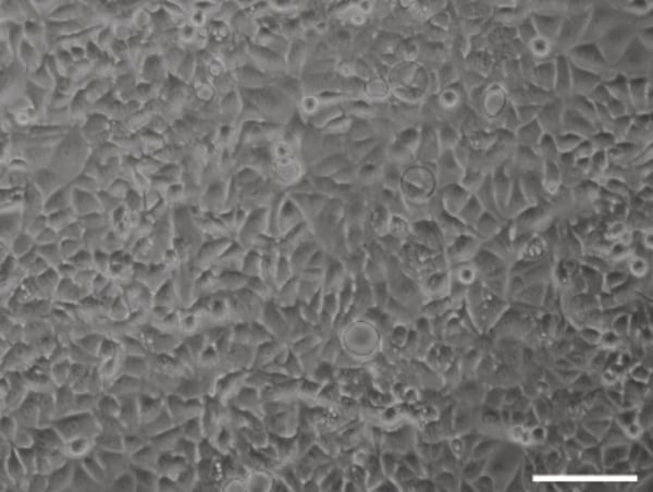
Figure 1. Phase contrast micrograph of MCF-7 cells grown in conventional 2D culture plates.
Scale bar: 200 µm.
- Complete media consisted of: Dulbecco’s Modified Eagles Medium (DMEM) supplemented with 10 % v/v FBS, 2 mM L-glutamine and 100 U/mL Penicillin/Streptomycin.
- Cells were harvested by trypsinisation and centrifuged for 5 minutes (1000 rpm). The supernatant was discarded and the cell pellet was re-suspended in an appropriate volume of media for cell-counting by Trypan Blue.
- Follow one of the following methods according to your choice of Alvetex Scaffold format.
2.2. 6 well insert format (AVP004)
- Cells were re-suspended at a concentration of 10 × 106 cells/mL for seeding.
- Alvetex Scaffold 6 well inserts were prepared for seeding with a 70 % ethanol wash (7 mL) and
subsequent media washes (twice, with 7 mL media each). - 100 μL of the cell suspension was added to the centre of the Alvetex Scaffold, which was
equivalent to 1 × 10⁶ cells per well. - The plate was incubated for 60 minutes at 37 °C with 5 % CO2 to allow the cells to settle into
the scaffold. - 10 mL of media was added to each well taking care not to dislodge cells from the membrane.
- Plates were re-incubated and maintained by complete media exchange after every 2-3 days.
Note: This method can be applied to the use of Alvetex Scaffold in12 well insert format (AVP005). Adjust cell seeding and media volumes according to the guidelines provided in the Alvetex Scaffold Quick Start protocol.
2.3. 24 well plate format (AVP006)
- Cells were re-suspended at a concentration of 4 × 10⁶ cells/mL for seeding.
- Alvetex Scaffold 24 well plate were prepared for seeding with a 70 % ethanol wash (2 mL per
well) and subsequent media washes (twice, with 2 mL media each). - 50 μL of the cell suspension was added to the centre of the Alvetex Scaffold, which was
equivalent to 0.2 × 10⁶ cells per well. - The plate was incubated for 60 minutes at 37 °C with 5 % CO2 to allow the cells to settle into
the scaffold. - 2 mL of media was added to each well taking care not to dislodge cells from the membrane.
- Plates were re-incubated and maintained by complete media exchange every 2-3 days for the
first 10 days, then every day.
Note: This method can be applied to the use of Alvetex Scaffold in 12 well insert format (AVP005). Adjust cell seeding and media volumes according to the guidelines provided in the Alvetex Scaffold Quick Start protocol.
3. Example data
3.1. 6 well insert format (AVP004)
MCF-7 cells were maintained on Alvetex Scaffold 6 well inserts for up to 21 days. Cultures underwent complete media change every 2-3 days. At specific time points, cultures were harvested and analysed by histological staining (Figure 2) and MTT viability assay (Figure 3) to monitor cell survival and proliferation within the scaffold. For histology and MTT assay protocols refer to Alvetex Protocols.
The cells showed good linear growth and expansion in 3D which plateaued between 14 and 21 days. We would not recommend growing cultures of MCF-7 cells beyond 14-18 days as when they reach confluency in 3D they can become over-crowded and the quality of the culture is reduced resulting in cell death.
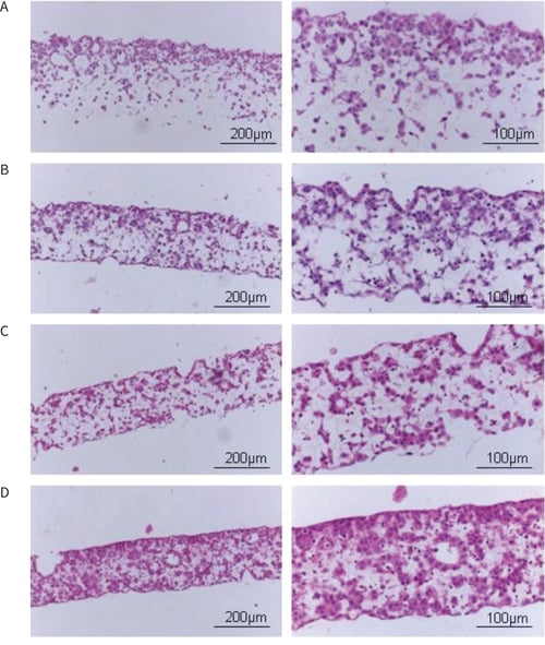
Figure 2. Brightfield micrographs showing the structure of MCF-7 cells cultured for (A) 3 days, (B) 7 days, (C) 10 days or (D) 14 days on 22 mm diameter Alvetex Scaffold presented in 6 well inserts. Cells were fixed, embedded in paraffin wax, sectioned (10 µm) and counterstained with haematoxylin and eosin.
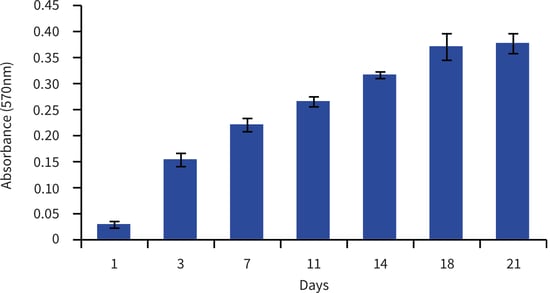
Figure 3. Biochemical analysis of cell viability using a standard MTT assay. Data from 3 sample replicates of MCF-7 cells are shown (mean ± SD). Each well was sampled in triplicate with mean value shown. Cells were cultured for up to 21 days on 22 mm Alvetex Scaffold presented in 6 well inserts. Absorbance = 570nm
3.2. 24 well plate format (AVP006)
MCF-7 cells were maintained on Alvetex Scaffold 24 well plates for up to 18 days. Cultures underwent complete media change every 2-3 days until day 10, then every day. At specific time- points, cultures were harvested and analysed by histological staining (Figure 2) and MTT viability assay (Figure 3) to monitor cell survival and proliferation within the scaffold. For histology and MTT assay protocols refer to Alvetex Protocols.
The cultures showed good linear growth and expansion in 3D which plateaued between 10 and 18 days. If cells are grown longer than 10 days, we would recommend changing the medium every day. When the cells reach confluency in 3D they become over-crowded and the quality of the culture is reduced, resulting in cell death, therefore we would not recommend culturing these cells for longer than 14 days in this culture format.
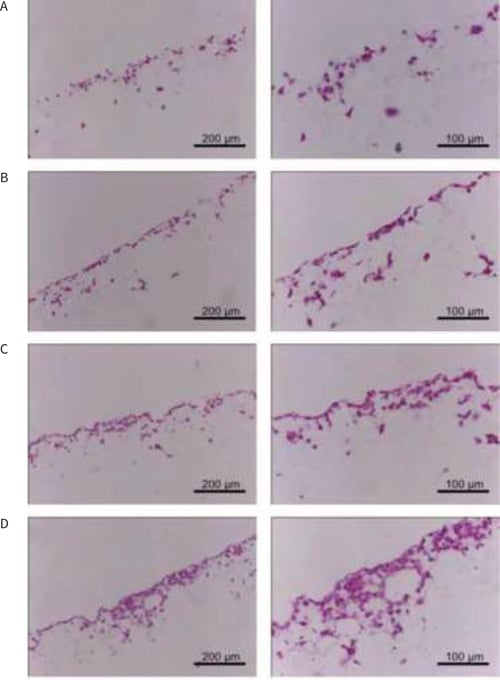
Figure 4. Brightfield micrographs showing the structure of MCF-7 cells cultured for (A) 3 days, (B) 7 days, (C) 10 days or (D) 14 days on 15 mm diameter Alvetex Scaffold presented in 24 well plates. Cells were fixed, embedded in paraffin wax, sectioned (10 µm) and counterstained with haematoxylin and eosin.
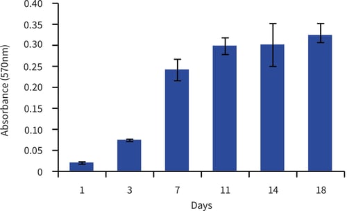
Figure 5. Biochemical analysis of cell viability using a standard MTT assay. Data from 3 sample replicates of MCF-7 cells are shown (mean ± SD). Each well was sampled in triplicate with mean value shown. Cells were cultured for up to 18 days on 15 mm Alvetex Scaffold presented in 24 well plate format.