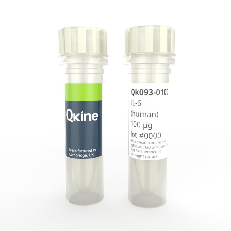
Recombinant human IL-6 protein
QK093
Brand: Qkine
Interleukin-6 (IL-6) is a multifunctional cytokine that regulates immune responses and inflammation. It is produced by various cells, including immune cells such as T cells and macrophages, as well as non-immune cells like fibroblasts and endothelial cells.
Human IL-6 has a molecular weight of 20.9 kDa. This protein is animal origin-free, carrier-free and tag-free to ensure its purity with exceptional lot-to-lot consistency. IL-6 is suitable for the culture of reproducible and high-quality hematopoietic stem cells and other relevant cells.

Currency:
| Product name | Catalog number | Pack size | Price | Price (USD) | Price (GBP) | Price (EUR) |
|---|---|---|---|---|---|---|
| Recombinant human IL-6 protein, 25 µg | QK093-0025 | 25 µg | (select above) | $ 340.00 | £ 250.00 | € 292.00 |
| Recombinant human IL-6 protein, 50 µg | QK093-0050 | 50 µg | (select above) | $ 520.00 | £ 380.00 | € 444.00 |
| Recombinant human IL-6 protein, 100 µg | QK093-0100 | 100 µg | (select above) | $ 685.00 | £ 500.00 | € 584.00 |
| Recombinant human IL-6 protein, 500 µg | QK093-0500 | 500 µg | (select above) | $ 2775.00 | £ 2025.00 | € 2366.00 |
| Recombinant human IL-6 protein, 1000 µg | QK093-1000 | 1000 µg | (select above) | $ 4340.00 | £ 3190.00 | € 3726.00 |
Note: prices shown do not include shipping and handling charges.
Qkine company name and logo are the property of Qkine Ltd. UK.
Alternative protein names
B-cell stimulatory factor 2 (BSF-2)
CTL differentiation factor (CDF)
Hybridoma growth factor
Interferon beta-2 (IFN-beta-2)
Species reactivity
human
species similarity:
mouse – 39%
rat – 38%
porcine – 59%
bovine – 55%
Summary
- High purity human IL-6 protein (Uniprot number: P05231)
- 20.9 kDa (monomer)
- >98%, by SDS-PAGE quantitative densitometry
- Expressed in E. coli
- Animal origin-free (AOF) and carrier protein-free
- Manufactured in Qkine's Cambridge, UK laboratories
- Lyophilized from HEPES, NaCl
- Resuspend in sterile-filtered water at >50 µg/ml, add carrier protein if desired, prepare single use aliquots and store frozen at -20 °C (short-term) or -80 °C (long-term).
Featured applications
- Stimulation of activated B cell proliferation
- Stimulation of T cell differentiation
- Hematopoietic stem cell regulation
- Proliferation and differentiation of erythrocytes, leukocytes and platelets
Bioactivity
IL-6 protein activity is determined using the IL-6-responsive firefly luciferase reporter assay. Transfected HEK293T cells are treated in triplicate with a serial dilution of IL-6 for 24 hours. Firefly luciferase activity is measured and normalised to the control Renilla luciferase activity. Data from Qk093 lot #204594. EC50 = 1.46 ng/mL (70 pM)

Purity
Recombinant IL-6 migrates as a major band at approximately 20 kDa in non-reducing (NR) and at approximately 18 kDa in reduced (R) conditions. No contaminating protein bands are present. The purified recombinant protein (3 µg) was resolved using 15% w/v SDS-PAGE in reduced (+β-mercaptoethanol, R) and non-reduced (NR) conditions and stained with Coomassie Brilliant Blue R250. Data from Qk093 lot #204599.

Further quality assays
- Mass spectrometry, single species with the expected mass
- Endotoxin: <0.005 EU/μg protein (below the level of detection)
- Recovery from stock vial: >95%

Qkine IL-6 is more biologically active than a comparable alternative supplier protein. Quantitative luciferase assay with Qkine IL-6 (Qk093, green) and alternative supplier IL-6 (Supplier B, black). Cells were treated in triplicate with a serial dilution of IL-6 for 24 hours. Firefly luciferase activity was measured and normalized to control Renilla luciferase activity. Qk093 EC50 1.85 ng/ml, Supplier B EC50 5.74 ng/ml.
Protein background
Interleukin-6 (IL-6) is a multifunctional cytokine that plays a crucial role in regulating the immune response, inflammation, and various physiological processes. IL-6 is produced by a variety of cells, including T cells, B cells, monocytes, fibroblasts, endothelial cells, and adipocytes [1].
IL6 protein is a pleiotropic cytokine that belongs to the interleukin family of proteins. IL-6 adopts a four-helix bundle structure, with helices A and D forming the receptor-binding site [2]. It is glycosylated, influencing stability and activity. IL-6 binds to IL-6 receptor (IL-6R), forming a hexameric complex with gp130, initiating downstream signaling via the JAK/STAT pathway. Conformational changes upon receptor binding facilitate signaling.
IL-6 has a primary involvement in the acute phase response, the immediate reaction to infection, injury, or inflammation. IL-6 stimulates the production of acute-phase proteins such as C-reactive protein (CRP), fibrinogen, and serum amyloid A, which help to enhance the immune response and facilitate tissue repair [3,4].
IL6 protein plays a key role in the regulation of the immune system. It promotes the differentiation of B cells into antibody-producing plasma cells and stimulates the proliferation and activation of T cells, enhancing the adaptive immune response. IL-6 acts on various immune cells to modulate inflammation, promoting the recruitment of immune cells to sites of infection or injury [5,6]. IL-6 has diverse effects on different tissues and organs throughout the body. It has been implicated in the regulation of metabolism, with studies suggesting that IL-6 may play a role in energy balance, glucose metabolism, and lipid metabolism. IL-6 has been shown to have both pro- and anti-inflammatory effects depending on the context and the cells involved [7].
Dysregulated IL-6 signaling has been associated with various pathological conditions, including autoimmune diseases, chronic inflammation, and cancer. Elevated levels of IL-6 have been observed in conditions such as rheumatoid arthritis, systemic lupus erythematosus, and inflammatory bowel disease, where it contributes to tissue damage and disease progression [8].
Background references
- Rose-John, S., & Heinrich, P. C. Soluble receptors for cytokines and growth factors: generation and biological function. Biochem. J. 436, 241–259 (2012).
- Kishimoto, T. Interleukin-6: Discovery of a pleiotropic cytokine. Arthritis Res. Ther. 12(Suppl 1), S2 (2010).
- Tanaka, T., Narazaki, M. & Kishimoto, T. Interleukin-6: From bench to bedside. Nat. Rev. Immunol. 20, 230–244 (2020).
- Scheller, J., Chalaris, A., Schmidt-Arras, D., & Rose-John, S. The pro- and anti-inflammatory properties of the cytokine interleukin-6. Biochim. Biophys. Acta – Mol. Cell Res. 1813, 878–888 (2011).
- Hunter, C. A., & Jones, S. A. IL-6 as a keystone cytokine in health and disease. Nat. Immunol. 16, 448–457 (2015).
- Rincon, M. Interleukin-6: from an inflammatory marker to a target for inflammatory diseases. Trends Immunol. 33, 571–577 (2012).
- Tanaka, T. & Kishimoto, T. The biology and medical implications of interleukin-6. Cancer Immunol. Res. 2, 288–294 (2014)
- Jones, S. A. & Jenkins, B. J. Recent insights into targeting the IL-6 cytokine family in inflammatory diseases and cancer. Nat. Rev. Immunol. 18, 773–789 (2018).