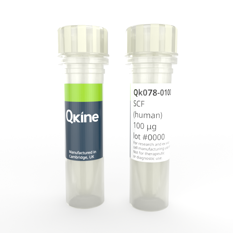
Recombinant human SCF protein
QK078
Brand: Qkine
Stem cell factor (SCF) is a critical factor in the maintenance and expansion of hematopoietic stem cells (HSCs) in the bone marrow microenvironment. Key myeloid progenitor differentiation factor for a variety of myeloid cells such as megakaryocytes, basophils, neutrophils, and monocytes, SCF is also a primary growth and activation factor for mast cells and eosinophils.
Recombinant SCF is a highly pure 18.5 kDa monomer, animal origin-free (AOF) and carrier-protein-free (CF). Qkine has optimized the SCF manufacture process to produce a highly bioactive protein with excellent lot-to-lot consistency for enhanced experimental reproducibility.

Currency:
| Product name | Catalog number | Pack size | Price | Price (USD) | Price (GBP) | Price (EUR) |
|---|---|---|---|---|---|---|
| Recombinant human SCF protein, 25 µg | QK078-0025 | 25 µg | (select above) | $ 260.00 | £ 190.00 | € 222.00 |
| Recombinant human SCF protein, 50 µg | QK078-0050 | 50 µg | (select above) | $ 380.00 | £ 280.00 | € 328.00 |
| Recombinant human SCF protein, 100 µg | QK078-0100 | 100 µg | (select above) | $ 560.00 | £ 410.00 | € 479.00 |
| Recombinant human SCF protein, 500 µg | QK078-0500 | 500 µg | (select above) | $ 2225.00 | £ 1625.00 | € 1898.00 |
| Recombinant human SCF protein, 1000 µg | QK078-1000 | 1000 µg | (select above) | $ 3465.00 | £ 2530.00 | € 2956.00 |
Note: prices shown do not include shipping and handling charges.
Qkine company name and logo are the property of Qkine Ltd. UK.
Alternative protein names
Stem cell growth factor(SCF)
C-kit ligand
Species reactivity
human
species similarity:
mouse – 79%
rat – 79%
porcine – 83%
bovine – 82%
Summary
- High purity human SCF protein (Uniprot number: P21583)
- 18.5 kDa (monomer)
- >98%, by SDS-PAGE quantitative densitometry
- Expressed in E. coli
- Animal origin-free (AOF) and carrier protein-free
- Manufactured in our Cambridge, UK laboratories
- Lyophilized from acetonitrile, TFA
- Resuspend in sterile-filtered water at >50 µg/ml, add carrier protein if desired, prepare single use aliquots and store frozen at -20 °C (short-term) or -80 °C (long-term).
Bioactivity
Recombinant SCF activity is determined using proliferation of TF-1 human myeloid leukemia cells. Cells are treated in triplicate with a serial dilution of SCF for 48 hours. Cell viability is measured using the CellTiter-Glo (Promega) luminescence assay. Data from Qk078 lot #204621. EC50 = 1.41 ng/mL (76 pM).

Purity
Recombinant SCF migrates at approximately 18 kDa (monomer) in reduced (R) and at approximately 15 kD in non-reduced (NR) conditions. No contaminating protein bands are present. The purified recombinant protein (3 µg) was resolved using 15% w/v SDS-PAGE in reduced (+β-mercaptoethanol, R) and non-reduced (NR) conditions and stained with Coomassie Brilliant Blue R250. Data from Qk078 lot #204621.

Further quality assays
- Mass spectrometry, single species with the expected mass
- Endotoxin: <0.005 EU/μg protein (below the level of detection)
- Recovery from stock vial: >95%

Qkine SCF has equivalent bioactivity to SCF from an alternative major supplier. Recombinant SCF activity was determined using proliferation of TF-1 human myeloid leukemia cells. Cells were treated in triplicate with a serial dilution of SCF for 48 hours. Cell viability was measured using the Cell Titer-Glo (Promega) luminescence assay. Data from Qk078 lot #204613. EC50 = Qk078: 2.86 ng/mL (154 pM). Supplier B: 3.05 ng/ml (166 pM)
Protein background
Stem Cell Factor (SCF), also known as kit-ligand or mast cell growth factor, is a pivotal protein in stem cell function, from embryonic development to tissue regeneration and cancer biology [1]. SCF functions primarily through interaction with its receptor, c-kit, a transmembrane tyrosine kinase receptor expressed on the surface of various cell types, including hematopoietic stem cells, neural crest-derived cells, melanocytes, and germ cells. Upon binding to c-kit, it triggers a cascade of intracellular signaling pathways, such as the Ras/MAPK, PI3K/Akt, and JAK/STAT pathways, which regulate cell proliferation, survival, migration, and differentiation [2,3].
Stem Cell Factor serves as a crucial ligand for its receptor, c-Kit, initiating signaling cascades upon binding. SCF can also be processed into a soluble form through proteolytic cleavage near the cell membrane. This cleavage releases the extracellular domain of SCF into the extracellular space, generating soluble SCF. Despite this modification, soluble SCF retains its ability to bind to c-Kit and activate downstream signaling pathways. Soluble SCF may act as a paracrine or autocrine factor, exerting its effects on nearby cells or the same cell producing it [3,4].
One of the key roles of SCF is in hematopoiesis, where SCF acts as a critical factor in the maintenance and expansion of hematopoietic stem cells (HSCs) in the bone marrow microenvironment [5,6]. It not only promotes the self-renewal of HSCs but also facilitates their differentiation into various blood cell lineages, including red blood cells, mast cells, and platelets [4].
Stem Cell Factor plays a crucial role in embryonic development, where it contributes to the formation of various tissues and organs. During embryogenesis, SCF is implicated in the proliferation, migration, and survival of neural crest cells, which give rise to a diverse array of cell types, including neurons, glial cells, melanocytes, and smooth muscle cells [7].
Beyond its roles in normal physiology, dysregulation of SCF signaling is associated with various pathological conditions, including cancer [8]. Aberrant activation of the SCF/c-kit pathway is observed in certain types of cancer, such as gastrointestinal stromal tumors (GISTs), acute myeloid leukemia (AML), and melanoma. In these malignancies, mutations in c-kit or overexpression of SCF contribute to uncontrolled cell proliferation, survival, and metastasis, making the SCF/c-kit axis an attractive target for cancer therapy [9].
Background references
- Huang EJ, Nocka KH, Buck J, Besmer P. Differential expression and processing of two cell associated forms of the kit-ligand: KL-1 and KL-2. Mol Biol Cell. 1992;3(3):349-362. https://doi.org/10.1091/mbc.3.3.349
- Lennartsson J, Rönnstrand L. Stem cell factor receptor/c-Kit: from basic science to clinical implications. Physiol Rev. 2012;92(4):1619-1649. https://doi.org/10.1152/physrev.00046.2011
- Yuzawa S, Opatowsky Y, Zhang Z, Mandiyan V, Lax I, Schlessinger J. Structural basis for activation of the receptor tyrosine kinase KIT by stem cell factor. Cell. 2007;130(2):323-334. doi: 10.1016/j.cell.2007.05.055
- Huang EJ, Nocka KH, Buck J, Besmer P. Differential expression and processing of two cell associated forms of the kit-ligand: KL-1 and KL-2. Mol Biol Cell. 1992;3(3):349-362. doi:10.1091/mbc.3.3.349
- Blume-Jensen P, Janknecht R, Hunter T. The kit receptor promotes cell survival via activation of PI 3-kinase and subsequent Akt-mediated phosphorylation of Bad on Ser136. Curr Biol. 1998;8(13):779-782. https://doi.org/10.1016/s0960-9822(98)70300-2
- Zsebo KM, Williams DA, Geissler EN, et al. Stem cell factor is encoded at the Sl locus of the mouse and is the ligand for the c-kit tyrosine kinase receptor. Cell. 1990;63(1):213-224. https://doi.org/10.1016/0092-8674(90)90303-v
- Waskow C, Madan V, Bartels S, et al. Hematopoietic stem cell transplantation without irradiation. Nat Methods. 2009;6(4):267-269. https://doi.org/10.1038/nmeth.1314
- Hu Y, Smyth GK. ELDA: extreme limiting dilution analysis for comparing depleted and enriched populations in stem cell and other assays. J Immunol Methods. 2009;347(1-2):70-78. https://doi.org/10.1016/j.jim.2009.06.008
- Linnekin D. Early signaling pathways activated by c-Kit in hematopoietic cells. Int J Biochem Cell Biol. 1999;31(10):1053-1074. https://doi.org/10.1016/s1357-2725(99)00076-0
Publications using recombinant human SCF protein (Qk078)
STAT3 signalling enhances tissue expansion during postimplantation mouse development
Azami T, Theeuwes B, Ton M-LN et al.
DOI: https://doi.org/10.1101/2024.10.11.617785
FAQ
What is Stem Cell Factor (SCF)?
SCF is a protein growth factor crucial for stem cell maintenance and differentiation, including hematopoietic stem cells in the bone marrow microenvironment.
Where is SCF found?
SCF is found in the bone marrow, skin, gastrointestinal tract, reproductive organs, and nervous system. It is produced by fibroblasts, endothelial cells, stromal cells, and epithelial cells to support tissue homeostasis and regulate cellular processes such as hematopoiesis, melanogenesis, and immune responses.
Is SCF a cytokine?
Yes, SCF is classified as a cytokine as it can be defined as a small protein that plays a crucial role in cell signalling and immune responses.
What does SCF bind to?
SCF binds to c-kit, which is also called the tyrosine-protein kinase kit or CD117. C-kit is a receptor tyrosine kinase that is expressed on the surface of hematopoietic stem cells, mast cells, melanocytes, germ cells, and interstitial cells.
What is the function of the SCF receptor?
Upon binding of SCF to c-kit, it triggers intracellular signaling cascades that regulate cell survival, proliferation, and differentiation.
What is the SCF pathway?
The SCF/c-kit signaling pathway.
How is SCF used in cell culture?
SCF is added to cell culture media as a supplement to provide essential signals for stem cell survival, proliferation, and differentiation. It acts as a growth factor, stimulating the activation of signaling pathways that regulate cellular processes crucial for stem cell function.