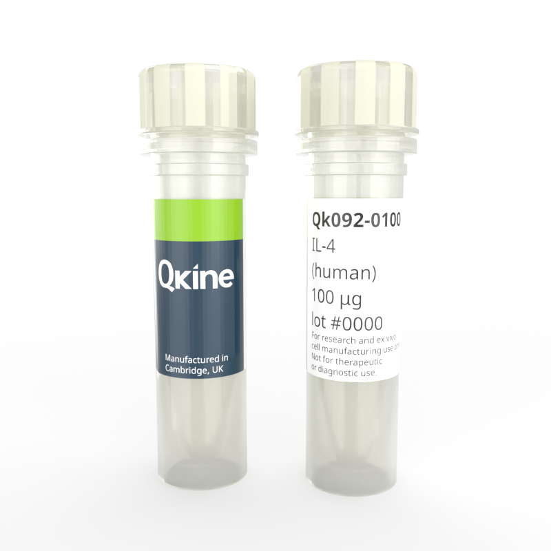
Recombinant human IL-4 protein
QK092
Brand: Qkine
Interleukin-4 (IL-4) is a pleiotropic, immune-modulatory cytokine that is secreted primarily by mast cells, T-cells, eosinophils, and basophils. IL-4 plays a crucial role in hematopoiesis, the regulation of antibody production, the stimulation of activated B cell and T cell proliferation, and the differentiation of B cells into plasma cells
Human IL-4 has a molecular weight of 15.1 kDa. This protein is animal origin-free, carrier-free and tag-free to ensure its purity with exceptional lot-to-lot consistency. Qk092 is suitable for the culture of reproducible and high-quality stem cells and other relevant cells.

Currency:
| Product name | Catalog number | Pack size | Price | Price (USD) | Price (GBP) | Price (EUR) |
|---|---|---|---|---|---|---|
| Recombinant human IL-4 protein, 25 µg | QK092-0025 | 25 µg | (select above) | $ 340.00 | £ 250.00 | € 292.00 |
| Recombinant human IL-4 protein, 50 µg | QK092-0050 | 50 µg | (select above) | $ 520.00 | £ 380.00 | € 444.00 |
| Recombinant human IL-4 protein, 100 µg | QK092-0100 | 100 µg | (select above) | $ 685.00 | £ 500.00 | € 584.00 |
| Recombinant human IL-4 protein, 500 µg | QK092-0500 | 500 µg | (select above) | $ 2775.00 | £ 2025.00 | € 2366.00 |
| Recombinant human IL-4 protein, 1000 µg | QK092-1000 | 1000 µg | (select above) | $ 4370.00 | £ 3190.00 | € 3726.00 |
Note: prices shown do not include shipping and handling charges.
Qkine company name and logo are the property of Qkine Ltd. UK.
Alternative protein names
B-cell stimulatory factor 1 (BSF-1)
Binetrakin
Lymphocyte stimulatory factor 1
Pitrakinra
Species reactivity
human
species similarity:
mouse – 39%
rat – 38%
porcine – 59%
bovine – 55%
Frequently used together:
Summary
- High purity human IL-4 protein (Uniprot number: P05112)
- 15.1 kDa (monomer)
- >98%, by SDS-PAGE quantitative densitometry
- Expressed in E. coli
- Animal origin-free (AOF) and carrier protein-free
- Manufactured in our Cambridge, UK laboratories
- Lyophilized from acetonitrile, TFA
- Resuspend in sterile-filtered water at >50 µg/ml, add carrier protein if desired, prepare single use aliquots and store frozen at -20 °C (short-term) or -80 °C (long-term).
Featured applications
- Stimulation of activated T cell proliferation
- Stimulation of activated B cell proliferation
- Differentiation of B cells into plasma cells
- Lymphoid differentiation
Bioactivity
IL-4 activity is determined using proliferation of TF-1 human myeloid leukemia cells. EC50 = 235 pg/mL (16 pM). Cells are treated in triplicate with a serial dilution of IL-4 for 72 hours. Cell viability is measured using the CellTiter-Glo (Promega) luminescence assay. Data from Qk092 lot #204598.

Purity
Recombinant IL-4 migrates as a major band at approximately 14 kDa in non-reducing (NR) and at approximately 12.5 kDa in reduced (R) conditions. No contaminating protein bands are present. The purified recombinant protein (3 µg) was resolved using 15% w/v SDS-PAGE in reduced (+β-mercaptoethanol, R) and non-reduced conditions and stained with Coomassie Brilliant Blue R250. Data from Qk092 batch #204598.

Further quality assays
- Mass spectrometry, single species with the expected mass
- Endotoxin: <0.005 EU/μg protein (below the level of detection)
- Recovery from stock vial: >95%

Qkine IL-4 is as biologically active as a comparable alternative supplier protein. Stimulation of proliferation of TF-1 cells with Qkine IL-4 (Qk092, green) and alternative supplier IL-4 (Supplier B, black). Cells were treated in triplicate with a serial dilution of IL-4 for 72 hours and proliferation measured using the CellTiter-Glo (Promega) luminescence assay.
Protein background
Interleukin 4 (IL-4) is an important signaling molecule within the immune system, playing multiple roles in orchestrating immune responses and maintaining immune homeostasis. IL-4 is primarily produced by immune cells, including mast cells, T helper 2 (Th2) cells, eosinophils, and basophils, and exerts its effects through interaction with its specific receptor, IL-4Rα and subsequent activation of downstream signaling pathways [1]. IL-4 plays a crucial role in hematopoiesis, the regulation of antibody production, the stimulation of activated B cell and T cell proliferation, and the differentiation of B cells into plasma cells. IL-4 induces the expression of class II MHC molecules on resting B-cells and aids regulation of the low-affinity Fc receptor for IgE (CD23) expression on lymphocytes and monocytes [2].
IL-4 has a compact, globular fold stabilized by three disulfide bonds. IL-4 consists of a characteristic four-alpha helix bundle with a left-handed twist, along with a two-stranded anti-parallel beta-sheet [2]. This structural arrangement allows stability to IL-4 and also facilitates its interaction with its receptor, IL-4Rα. The receptor for IL-4, IL-4Rα, exists in three distinct complexes within the body, each playing a crucial role in mediating IL-4 signaling [3]. Type 1 receptors consist of the IL-4Rα subunit coupled with a common gamma chain, while type 2 receptors comprise the IL-4Rα subunit bound to IL-13Rα1. Type 1 receptors specifically bind IL-4, whereas type 2 receptors have the capacity to bind both IL-4 and IL-13, two cytokines with closely related functions. This differential receptor composition allows for nuanced regulation of immune responses depending on the specific ligands present [4].
IL-4 serves as a regulator of immune cell differentiation and activation [5]. It promotes the differentiation of naive T cells into Th2 cells, which are crucial for orchestrating immune responses against extracellular pathogens and for mediating allergic reactions. Additionally, IL-4 stimulates the proliferation and activation of B cells, leading to the production of antibodies and the formation of memory B cells [6]. IL-4 plays a pivotal role in modulating macrophage polarization and the development of an alternative activation phenotype (M2) associated with tissue repair and resolution of inflammation [7].
In the context of disease, dysregulation of IL-4 signaling has been implicated in various immune disorders, including allergies, asthma, and autoimmune diseases. Enhanced IL-4 production or aberrant IL-4 receptor signaling can contribute to the pathogenesis of allergic inflammation and tissue damage. Therapeutic strategies aimed at targeting IL-4 or its receptor have shown promise in treating these conditions by modulating immune responses and dampening inflammation [8].
Background references
- Paul, W. E. Interleukin 4: a prototypic immunoregulatory lymphokine. Blood 77, 1859–1870 (1991). PMID: 2018830
- Gadani, S. P., Cronk, J. C., Norris, G. T., & Kipnis, J. IL-4 in the brain: a cytokine to remember. Journal of Immunology 189, 4213–4219 (2012). doi: 10.4049/jimmunol.1202246
- Nelms, K., Keegan, A. D., Zamorano, J., Ryan, J. J., & Paul, W. E. The IL-4 receptor: signaling mechanisms and biologic functions. Annual Review of Immunology 17, 701–738 (1999). doi: 10.1146/annurev.immunol.17.1.701
- Ul-Haq, Z., Naz, S., & Mesaik, M. A. Interleukin-4 receptor signaling and its binding mechanism: A therapeutic insight from inhibitors tool box. Cytokine & Growth Factor Reviews 32, 3–15 (2016). doi: 10.1016/j.cytogfr.2016.04.002
- Gordon, S., & Martinez, F. O. Alternative activation of macrophages: mechanism and functions. Immunity 32, 593–604 (2010). doi: 10.1016/j.immuni.2010.05.007.
- Bhattarai, P et al. IL4/STAT6 Signaling Activates Neural Stem Cell Proliferation and Neurogenesis upon Amyloid-β42 Aggregation in Adult Zebrafish Brain. Cell Reports 17, 941–948 (2016). doi: 10.1016/j.celrep.2016.09.075
- Hershey, G. K. IL-13 receptors and signaling pathways: an evolving web. Journal of Allergy and Clinical Immunology 111, 677–690 (2003). doi: 10.1067/mai.2003.1333
- Chatila, T. A. Interleukin-4 receptor signaling pathways in asthma pathogenesis. Trends in Molecular Medicine 10, 493–499 (2004). doi: 10.1016/j.molmed.2004.08.004