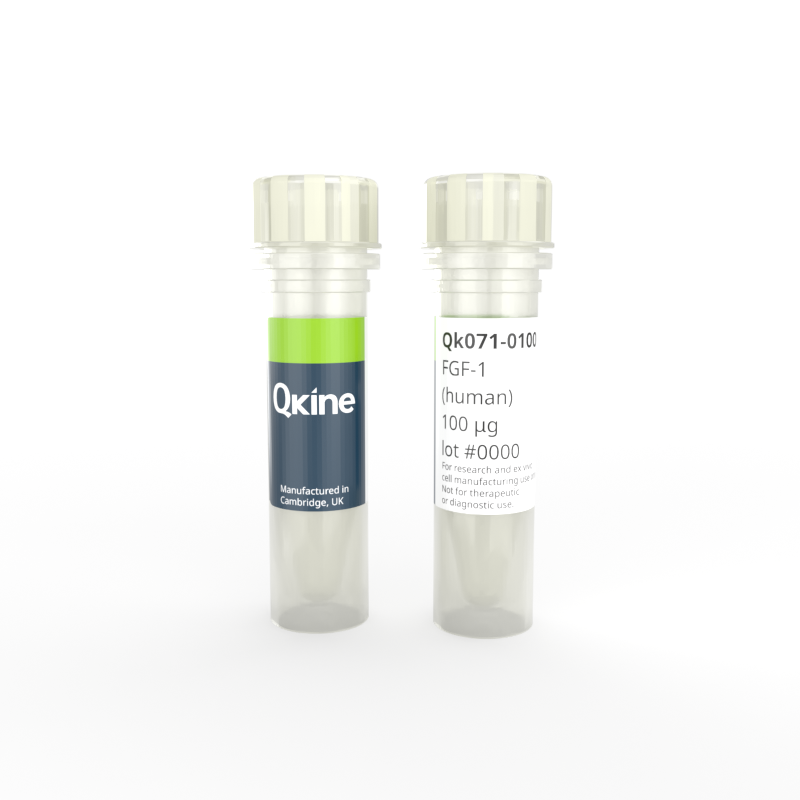
Recombinant human FGF-1 protein
QK071
Brand: Qkine
Fibroblast Growth Factor 1 (FGF-1) can stimulate growth and differentiation of endothelial and epithelial cells and the development of organoids. FGF-1 can also be used for the maintenance of oligodendrocytes and astroglia as well as bone marrow-derived mesenchymal and hematopoietic stem cells.
Qkine human FGF-1 has a molecular weight of 15.9 kDa. This protein is animal origin-free, carrier-free and tag-free to ensure its purity with exceptional lot-to-lot consistency. Qk071 is suitable for the culture of reproducible mesenchymal, endothelial, hematopoietic, glial, and other relevant cells.

Currency:
| Product name | Catalog number | Pack size | Price | Price (USD) | Price (GBP) | Price (EUR) |
|---|---|---|---|---|---|---|
| Recombinant human FGF-1 protein, 50 µg | QK071-0050 | 50 µg | (select above) | $ 240.00 | £ 175.00 | € 205.00 |
| Recombinant human FGF-1 protein, 100 µg | QK071-0100 | 100 µg | (select above) | $ 315.00 | £ 230.00 | € 269.00 |
| Recombinant human FGF-1 protein, 500 µg | QK071-0500 | 500 µg | (select above) | $ 810.00 | £ 590.00 | € 690.00 |
| Recombinant human FGF-1 protein, 1000 µg | QK071-1000 | 1000 µg | (select above) | $ 1205.00 | £ 880.00 | € 1028.00 |
Note: prices shown do not include shipping and handling charges.
Qkine company name and logo are the property of Qkine Ltd. UK.
Alternative protein names
Species reactivity
human
species similarity:
mouse – 96%
rat – 96%
porcine – 97%
bovine – 92%
Summary
- High purity human protein (Uniprot number: P05230)
- 15.9 kDa (monomer)
- >98%, by SDS-PAGE quantitative densitometry
- Expressed in E. coli
- Animal origin-free (AOF) and carrier protein-free
- Manufactured in our Cambridge, UK laboratories
- Lyophilized from Tris, NaCl, CyS
- Resuspend in sterile-filtered water at >50 µg/ml, add carrier protein if desired, prepare single use aliquots and store frozen at -20 °C (short-term) or -80 °C (long-term).
Featured applications
- Epithelial organoid culture
- Differentiation of iPSCs into epithelial cells
- Mesenchymal stem cell research
- Organoid growth and proliferation
- Differentiation of neural progenitor cells into oligodendrocyte precursor cells
- Maintenance and differentiation of hematopoietic stem cells
Bioactivity
FGF-1 activity was determined using the FGF-1-responsive firefly luciferase reporter assay. HEK293T cells were treated in triplicate with a serial dilution of FGF-1 for 3 hours. Firefly luciferase activity was measured and normalised to the control Renilla luciferase activity. EC50 = 0.81 ng/ml (51 pM). Data from Qk071 lot #204543.

Purity
Recombinant FGF-1 migrates as a major band at approximately 15.97 kDa in non-reduced (NR) and reduced (R) conditions. No contaminating protein bands are present. The purified recombinant protein (3 µg) was resolved using 15% w/v SDS-PAGE in reduced (+β-mercaptoethanol, R) and non-reduced (NR) conditions and stained with Coomassie Brilliant Blue R250. Data from Qk071 batch #204543.

Further quality assays
- Mass spectrometry, single species with the expected mass
- Endotoxin: <0.005 EU/μg protein (below the level of detection)
- Recovery from stock vial: >95%

Recombinant FGF-1 migrates as a major band at approximately 15.97 kDa in non-reduced (NR) and reduced (R) conditions. No contaminating protein bands are present. The purified recombinant protein (3 µg) was resolved using 15% w/v SDS-PAGE in reduced (+β-mercaptoethanol, R) and non-reduced (NR) conditions and stained with Coomassie Brilliant Blue R250. Data from Qk071 batch #204543.
Protein background
Fibroblast Growth Factor 1 (FGF-1) is a member of the FGF family and regulates the proliferation, migration, and differentiation of mesenchymal cells [1–3]. It plays a crucial role in multiple biological processes including embryonic development and tissue regeneration [1,2,4,5]. It is a key regulator of angiogenesis and wound healing as it regulates the proliferation and maintenance of endothelial and epithelial cells [3]. It has neurotrophic properties to protect and repair neurons and lipid metabolism functions to regulate adipocytes [5,6]. FGF-1 can also promote the differentiation of hematopoietic stem cells [7]. Notably, FGF-1 is implicated in the tumour growth and migration [8].
FGF-1 is composed of 155 amino acids, with a molecular weight of approximately 17-18 kDa [2,9]. It consists of 12 anti-parallel β-strands organized into a three-fold symmetric β-sheet [10]. FGF-1 binds to different FGF receptors such as FGFR1 triggering several signaling cascades involved in cell growth, proliferation, migration, survival, and differentiation. These include the Ras/Raf/Mek/Erk, Pi3k/Akt, Jnk/Mapk, and STAT3/Nf-kb pathways [8].
The role of FGF-1 on embryonic development and the regulation of mesenchymal cells makes it a growth factor used for a range of different cultures in vitro. FGF-1 is used to promote the differentiation and proliferation of endothelial cells and epithelial cells [11,12]. As FGF-1 also promotes the branching of epithelial cells, it has been used for embryonic lung epithelium cultures and human iPSC-derived uretic bud organoids13,14. Additionally, its neurotrophic properties make it ideal for the maintenance of neural progenitors as well as supporting cells such as oligodendrocytes, and astroglia [5,15–17]. FGF-1 has also been reported for the culture of bone marrow-derived mesenchymal and hematopoietic stem and progenitor cells [7,18,19].
Because of its diverse roles in cellular processes, FGF-1 is a target of interest in various clinical applications, including regenerative medicine, wound healing therapies, and potential treatments for metabolic disorders and neurodegenerative diseases [3,6,16]. In Type 2 diabetes, FGF-1 injections could lower the glucose level without risk of hypoglycaemia through its effect on glucose-sensing neuronal circuits [6]. In neurodegenerative diseases such as multiple sclerosis, FGF-1 could promote the remyelination of neurons [16]. Its role in angiogenesis could have great potential for novel therapies for myocardial infarction4. Finally, its involvement in cancer progression has led to investigations into targeted therapies to inhibit FGF-1 signaling in cancer cells.
Background references
- FGF1 fibroblast growth factor 1 [Homo sapiens (human)] - Gene - NCBI. https://www.ncbi.nlm.nih.gov/gene/2246.
- Ornitz, D. M. and Itoh, N. Fibroblast growth factors. Genome Biol. 2, reviews3005.1 (2001). doi: 10.1186/gb-2001-2-3-reviews3005
- Zakrzewska, M., Marcinkowska, E. and Wiedlocha, A. FGF-1: From Biology Through Engineering to Potential Medical Applications. Crit. Rev. Clin. Lab. Sci. 45, 91–135 (2008). doi: 10.1080/10408360701713120
- Engel, F. B., Hsieh, P. C. H., Lee, R. T. and Keating, M. T. FGF1/p38 MAP kinase inhibitor therapy induces cardiomyocyte mitosis, reduces scarring, and rescues function after myocardial infarction. Proc. Natl. Acad. Sci. 103, 15546–15551 (2006). doi: 10.1073/pnas.0607382103
- Nurcombe, V., Ford, M. D., Wildschut, J. A. and Bartlett, P. F. Developmental Regulation of Neural Response to FGF-1 and FGF-2 by Heparan Sulfate Proteoglycan. Science 260, 103–106 (1993). doi: 10.1126/science.7682010
- Gasser, E., Moutos, C. P., Downes, M. and Evans, R. M. FGF1 — a new weapon to control type 2 diabetes mellitus. Nat. Rev. Endocrinol. 13, 599–609 (2017). doi: 10.1038/nrendo.2017.78
- Walenda, T. et al. Synergistic effects of growth factors and mesenchymal stromal cells for expansion of hematopoietic stem and progenitor cells. Exp. Hematol. 39, 617–628 (2011). doi: 10.1016/j.exphem.2011.02.011
- Raju, R. et al. A Network Map of FGF-1/FGFR Signaling System. J. Signal Transduct. 2014, 962962 (2014). doi: 10.1155/2014/962962
- Liu, Y. et al. Advances in FGFs for diabetes care applications. Life Sci. 310, 121015 (2022). doi: 10.1016/j.lfs.2022.121015
- Zhu, X. et al. Three-Dimensional Structures of Acidic and Basic Fibroblast Growth Factors. Science 251, 90–93 (1991). doi: 10.1126/science.1702556
- Kang, S. S., Gosselin, C., Ren, D. & Greisler, H. P. Selective stimulation of endothelial cell proliferation with inhibition of smooth muscle cell proliferation by fibroblast growth factor-1 plus heparin delivered from fibrin glue suspensions. Surgery 118, 280–287 (1995). doi: 10.1016/s0039-6060(05)80335-6
- Ramos, C. et al. FGF-1 reverts epithelial-mesenchymal transition induced by TGF-β1 through MAPK/ERK kinase pathway. Am. J. Physiol.-Lung Cell. Mol. Physiol. 299, L222–L231 (2010). doi: 10.1152/ajplung.00070.2010
- Cardoso, W. V., Itoh, A., Nogawa, H., Mason, I. and Brody, J. S. FGF-1 and FGF-7 induce distinct patterns of growth and differentiation in embryonic lung epithelium. Dev. Dyn. 208, 398–405 (1997). doi: 10.1002/(SICI)1097-0177(199703)208:3<398::AID-AJA10>3.0.CO;2-X
- Mae, S.-I. et al. Expansion of Human iPSC-Derived Ureteric Bud Organoids with Repeated Branching Potential. Cell Rep. 32, 107963 (2020). doi: 10.1016/j.celrep.2020.107963
- Jiang, P. and Deng, W. Regenerating white matter using human iPSC-derived immature astroglia. Neurogenesis 3, e1224453 (2016). doi: 10.1080/23262133.2016.1224453
- Mohan, H. et al. Transcript profiling of different types of multiple sclerosis lesions yields FGF1 as a promoter of remyelination. Acta Neuropathol. Commun. 2, 178 (2014). doi: 10.1186/s40478-014-0168-9
- Jiang, P. et al. Human iPSC-Derived Immature Astroglia Promote Oligodendrogenesis by Increasing TIMP-1 Secretion. Cell Rep. 15, 1303–1315 (2016). doi: 10.1016/j.celrep.2016.04.011
- Haan, G. de et al. In Vitro Generation of Long-Term Repopulating Hematopoietic Stem Cells by Fibroblast Growth Factor-1. Dev. Cell 4, 241–251 (2003). doi: 10.1016/s1534-5807(03)00018-2
- Jiang, S. et al. Novel insights into a treatment for aplastic anemia based on the advanced proliferation of bone marrow‑derived mesenchymal stem cells induced by fibroblast growth factor 1. Mol. Med. Rep. 12, 7877–7882 (2015). doi: 10.3892/mmr.2015.442