Alvetex Scaffold Application Note 10
Enhanced Structure and Viability of Liver Hepatocytes
Download this application note as a PDF (2.39 MB)
Alvetex Scaffold is an ideal 3D substrate for in vitro culture, resulting 3D growth and extensive cell-cell contact for a wide variety of cell types.
Hepatocytes constitute the major metabolic cell type of the liver, and are central to in vitro assays designed to investigate the metabolism, biotransformation and toxicology of prospective pharmaceuticals. Liver cell lines and primary hepatocytes can be used to predict biological responses to xenobiotics in vitro.
Cell lines typically lack true differentiated functionality required to reflect hepatocytes in vivo, and while primary hepatocytes are the most representative liver cell type, once removed from their native tissue environment their viability and functionality rapidly decline. The re-creation of 3D tissue environments in vitro holds real promise for the development of more representative cell based assays, and modern progress in the development of novel 3D cell culture platforms has been rapid. Alvetex Scaffold technology is an ideal basis for drug toxicology assay development due to its ease of use, its inert and reproducible structure, and its suitability for adaptation into common cell culture formats. In this short application note, we demonstrate the growth patterns and behaviour of various liver-derived cell types including common cell lines and primary hepatocytes. The data presented include morphological analysis of cell growth by histology, and assessment of metabolic viability and functionality were examined using a range of common assays. Liver cells cultured in Alvetex Scaffold are morphologically and functionally superior to counterparts cultured by conventional 2D methods. Alvetex Scaffold presents itself as a platform from which superior in vitro assays can be developed, for use in both basic research and industrial settings.
Highlights
- Enhanced morphology and cellular structure of liver cells
- Increased viability and hepatic functionality
- Enhanced liver cell function by hepatocyte and endothelial cell co-culture
- Enhanced basal and inducible levels of phase 1 metabolic enzymes
Key Benefits
- Improved cell-cell interaction and development potential
- Functional advantage over 2D culture for improved in vitro hepatocyte models
- Platform for improved in vitro drug metabolism and toxicity assays
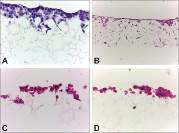
Figure 1. 3D culture of hepatocytes from different sources. The 3D growth of hepatocytes from various sources including cell lines and primary cells have been tested on Alvetex Scaffold. Brightfield images show examples of four different hepatocyte cell types. Cell cultures were preserved in Bouinʼs fixative, paraffin embedded, sectioned (10 μm) and counterstained with Haematoxylin and Eosin according to the protocol (see protocols). (A) HepG2 cells (ATCC, HB-8065) cultured in Alvetex Scaffold in 6-well insert (AVP004) format, 40× objective; (B) Upcyte hepatocytes (Medicyte GmbH) cultured in 24-well plate (AVP006) format, 20× objective; (C) Cryo-preserved rat hepatocytes (Biopredic Int.) cultured in 12-well insert (AVP005) format, 40× objective; (D) Rat hepatocytes (Abcellute Ltd.) cultured in 12-well insert (AVP005) format, 40× objective.
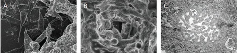
Figure 2. HepG2 form 3D structures when grown in Alvetex Scaffold. The hepatic cell line HepG2 shows improved growth and structure when grown in 3D culture compared to conventional 2D methods. Scanning electron micrographs of HepG2 cells cultured for 7 days in 2D (A) and in 3D in Alvetex Scaffold (B), showing increased homogeneity and enhanced cell-cell interactions and development of complex structures such as microvilli in the scaffold. High magnification transmission electron micrograph (C) of HepG2 cells cultured for 21 days in 3D in Alvetex Scaffold, showing the formation of microvilli, tight junctions, and bile canaliculi-like structures (taken from [1]).
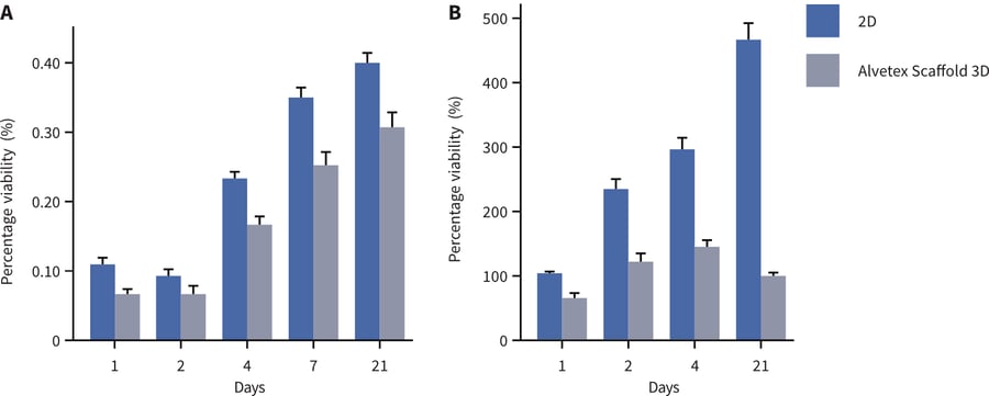
Figure 3. HepG2 cells show greater viability and albumin production when grown as 3D cultures using Alvetex Scaffold. Analysis of HepG2 cells using a standard MTT viability assay (A) demonstrated enhanced viability in Alvetex Scaffold over a period of 21 days in culture compared with cells grown in 2D culture. Similarly, albumin secretion into culture media (B) was greater in 3D Alvetex Scaffold cultures than in 2D counterparts (taken from [2]).
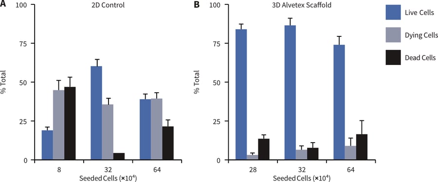
Figure 4. Enhanced viability of primary rat hepatocytes grown on Alvetex Scaffold. Viability of rat primary hepatocytes was determined by calcein AM / ethidium heterodimer labelling and confocal microscopy. Cells were seeded at different densities (8×, 32×, and 64× 10⁴ cells per well) cultured for 24 hours in 2D (A) and in 3D in Alvetex Scaffold (B). Cell viability in 2D was severely compromised at the lowest seeding density (10 % viability) and increasing cell seeding density resulted in comparable numbers of viable versus dead/dying cells. The highest proportion of viable cells achievable in 2D was 57 %. In 3D culture in Alvetex Scaffold, were highly viable (> 74 %), independent of the seeding density (taken from [3]).
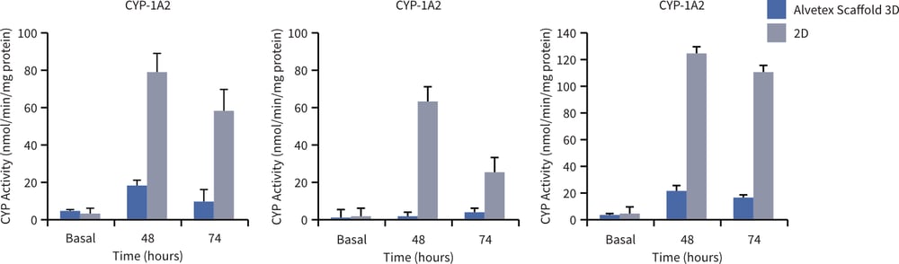
Figure 5. Induction of CYP enzyme expression is increased in rat primary hepatocytes grown in 3D culture using Alvetex Scaffold. Rat hepatocytes were cultured in 2D (grey bars) and 3D in Alvetex Scaffold (black bars) for 48 or 72 hours after induction and assay of different CYP isoforms. Basal enzyme activities are also shown. Culture in Alvetex Scaffold resulted in enhanced inducible levels of all three enzymes tested (CYP-1A2, -2B1, -3A2) (taken from [3]).
References
- Bokhari M, et al. 2007. Culture of HepG2 liver cells on three-dimensional polystyrene scaffolds enhances cell structure and function during toxicological challenge. J Anat. 211(4): 567-76.
- Bokhari M, et al. 2007. Novel cell culture device enabling three-dimensional cell growth and improved cell function. Biochem Biophys Res Commun. 354(4): 1095-100.
- Schutte M, et al. 2011. Rat primary hepatocytes show enhanced performance and sensitivity to acetaminophen during three-dimensional culture on a polystyrene scaffold designed for routine use. Assay Drug Dev Technol. 9(5): 475-86.