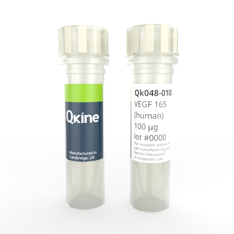
Recombinant human VEGF 165 protein
QK048
Brand: Qkine
Recombinant human vascular endothelial growth factor 165 (VEGF165/ VEGF-165/ VEGF165) protein is widely used in culturing primary endothelial cells, such as human umbilical vein endothelial cells (HUVEC).
VEGF 165 is commonly used with human-induced pluripotent stem cells or embryonic stem cells-derived endothelial cells for developing human vascular tissue models. It has many applications including its use in neural research involving oligodendrocyte precursor cells, Schwann cells, astrocytes, and microglia. It plays a role in bone formation, regulates mesenchymal stem cell differentiation, and serves as a survival factor for chondrocytes, hematopoietic stem cells, and tumor cells.

Currency:
| Product name | Catalog number | Pack size | Price | Price (USD) | Price (GBP) | Price (EUR) |
|---|---|---|---|---|---|---|
| Recombinant human VEGF 165 protein, 25 µg | QK048-0025 | 25 µg | (select above) | $ 340.00 | £ 250.00 | € 292.00 |
| Recombinant human VEGF 165 protein, 50 µg | QK048-0050 | 50 µg | (select above) | $ 520.00 | £ 380.00 | € 444.00 |
| Recombinant human VEGF 165 protein, 100 µg | QK048-0100 | 100 µg | (select above) | $ 685.00 | £ 500.00 | € 584.00 |
| Recombinant human VEGF 165 protein, 500 µg | QK048-0500 | 500 µg | (select above) | $ 2775.00 | £ 2025.00 | € 2366.00 |
| Recombinant human VEGF 165 protein, 1000 µg | QK048-1000 | 1000 µg | (select above) | $ 4370.00 | £ 3190.00 | € 3726.00 |
Note: prices shown do not include shipping and handling charges.
Qkine company name and logo are the property of Qkine Ltd. UK.
Alternative protein names
Species reactivity
human
species similarity:
mouse – 88%
rat – 88%
porcine – 97%
bovine – 94%
Summary
- High purity human VEGF 165 (Uniprot: P15692)
- 38.3 kDa (dimer), 19 kDa (monomer)
- >98%, by SDS-PAGE quantitative densitometry
- Expressed in E. coli
- Animal origin-free (AOF) and carrier protein-free
- Manufactured in Qkine'sCambridge, UK laboratories
- Lyophilized from acetonitrile, TFA
- Resuspend in sterile-filtered water at >50 µg/ml, add carrier protein if desired, prepare single use aliquots and store frozen at -20 °C (short-term) or -80 °C (long-term).
Featured applications
- Angiogenic cell research
- Endothelial cell differentiation
- iPSC-derived mesoderm differentiation
- Vasculature in organoids
- Neural stem cell research
- Mesenchymal stem cell research
Bioactivity
The bioactivity of Qk048 is measured using a luciferase reporter cell line which stably expresses the KDR (VEGFR-2) receptor. Cells were incubated with different concentrations of VEGF 165 for 6 hours before assaying for luciferase production. EC50=0.55 ng/ml (14.4 pM), data from Qk048 lot #104393, n=3.

Purity
Human VEGF 165 (Qk048) migrates as a dimer at 38 kDa in non-reducing (NR) conditions and as a monomer at 19 kDa upon reduction (R). No contaminating bands are visible. Purified recombinant protein (3 µg) was resolved using 15% w/v SDS-PAGE in reduced (+β-mercaptoethanol, R) and non-reduced (-β-mercaptoethanol, NR) conditions and stained with Coomassie Brilliant Blue R-250. Data from Qk048 lot #104393.

Further quality assays
- Mass spectrometry: single species with expected mass
- Endotoxin: <0.005 EU/μg protein (below level of detection)
- Recovery from stock vial: >95%

Qkine VEGF 165 has higher bioactivity than an alternative supplier. The bioactivity of Qkine and an alternative supplier were compared directly using a luciferase reporter cell line which stably expresses the KDR (VEGFR-2) receptor. Cells were incubated with different concentrations of VEGF 165 for 6 hours before assaying for luciferase production. EC50 = 0.55 ng/ml (14.4 pM) for Qk048, data from Qk048 lot #104393, n=3. EC50 = 1.44 ng/ml (37.6 pM) for Supplier 1 VEGF 165, , n=3.
Protein background
Vascular endothelial growth factor (VEGF) is a member of the platelet-derived growth factor (PDGF) family and a core regulator of angiogenesis and vascular permeability [1,2]. It is responsible for the survival, proliferation, migration, and specialization of endothelial cells, thus called an endothelial cell surviving factor [2,3].
In humans, VEGF is produced as multiple alternately spliced isoforms indicating the number of amino acids in length: VEGF121, VEGF145, VEGF148, VEGF162, VEGF165a, VEGF165b, VEGF183, VEGF189, and VEGF206. VEGF165 (or VEGF165a) is the most abundantly expressed isoform composed of 165 amino acids [1].
VEGF165 signals through the type I transmembrane receptor tyrosine kinases VEGFR1 (also called Flt-1) and VEGFR2 (Flk-1/KDR) [2]. Some VEGF family members (including VEGFA 165) also bind to the co-receptors neuropilin 1 (NRP1) and neuropilin 2 (NRP2), which can stimulate VEGFR2 activation. It is characterized by the presence of eight conserved cysteine residues forming a receptor-binding cystine-knot structure [4,5].
VEGF is widely used in culturing primary endothelial cells, such as human umbilical vein endothelial cells, under serum-free conditions for blood vessel developmental studies. VEGF165 is commonly used with human-induced pluripotent stem cells or embryonic stem cells-derived endothelial cells for developing human vascular tissue models for disease mechanism studies. Immunocytochemical staining with CD31 expression marker is used to indicate the presence of endothelial cells, thereby suggesting successful endothelial cell differentiation [6,7]. Endothelial cells can also be derived from hair follicle stem cells [8].
In addition to its angiogenic role, it is also involved in promoting neurogenesis and stimulating neural stem cell proliferation [9,10]. It can promote the proliferation, survival or migration of other glial cells such as oligodendrocyte precursor cells, Schwann cells, and can stimulate the expression of trophic factors by astrocytes [11]. Moreover, it is a critical factor in generating human pluripotent stem cell-derived vascularized brain organoids [6].
Finally, VEGF also plays a role in bone formation and regulates mesenchymal stem cell differentiation of pluripotent stem cells and bone marrow stem cells [12,13]. Therefore, it serves as a survival factor for chondrocytes, hematopoietic stem cells, and tumor cells [12,14].
The strong implications of VEGF have led to preclinical studies on the potential of VEGF administration in neurodegenerative and ischemic diseases [1,2,11]. The inhibition of VEGF has been approved for the treatment of neovascular ocular disease to prevent the blood brain barrier breakdown or excessive angiogenesis11. It is also a target in anti-angiogenic strategies in cancer due to its contribution in tumor angiogenesis [2,14].
Background references
- Bhisitkul, R. B. Vascular endothelial growth factor biology: clinical implications for ocular treatments. Br. J. Ophthalmol. 90, 1542–1547 (2006). doi: 10.1136/bjo.2006.098426
- Shibuya, M. Vascular Endothelial Growth Factor (VEGF) and Its Receptor (VEGFR) Signaling in Angiogenesis. Genes Cancer 2, 1097–1105 (2011). doi: 10.1177/1947601911423031
- Honnegowda, T. M., Kumar, P. & Rao, P. Role of angiogenesis and angiogenic factors in acute and chronic wound healing. Plast. Aesthetic Res. 2, null (2015). doi: 10.4103/2347-9264.165438
- Ferrara, N., Gerber, H.-P. & LeCouter, J. The biology of VEGF and its receptors. Nat. Med. 9, 669–676 (2003). doi.org/10.1038/nm0603-669
- Sulpice, E. et al. Neuropilin-1 and neuropilin-2 act as coreceptors, potentiating proangiogenic activity. Blood 111, 2036–2045 (2008). doi: 10.1182/blood-2007-04-084269
- Pham, M. T. et al. Generation of human vascularized brain organoids. Neuroreport 29, 588–593 (2018). doi: 10.1097/WNR.0000000000001014
- Atchison, L. et al. iPSC-Derived Endothelial Cells Affect Vascular Function in a Tissue-Engineered Blood Vessel Model of Hutchinson-Gilford Progeria Syndrome. Stem Cell Rep. 14, 325–337 (2020). doi: 10.1016/j.stemcr.2020.01.005
- Quan, R. et al. VEGF165 induces differentiation of hair follicle stem cells into endothelial cells and plays a role in in vivo angiogenesis. J. Cell. Mol. Med. 21, 1593–1604 (2017). doi: 10.1111/jcmm.13089
- Schänzer, A. et al. Direct Stimulation of Adult Neural Stem Cells In Vitro and Neurogenesis In Vivo by Vascular Endothelial Growth Factor. Brain Pathol. 14, 237–248 (2004). doi: 10.1111/j.1750-3639.2004.tb00060.x
- Storkebaum, E., Lambrechts, D. & Carmeliet, P. VEGF: once regarded as a specific angiogenic factor, now implicated in neuroprotection. BioEssays 26, 943–954 (2004). doi: 10.1002/bies.20092
- Lange, C., Storkebaum, E., de Almodóvar, C. R., Dewerchin, M. & Carmeliet, P. Vascular endothelial growth factor: a neurovascular target in neurological diseases. Nat. Rev. Neurol. 12, 439–454 (2016). doi: 10.1038/nrneurol.2016.88
- Berendsen, A. D. & Olsen, B. R. How Vascular Endothelial Growth Factor-A (VEGF) Regulates Differentiation of Mesenchymal Stem Cells. J. Histochem. Cytochem. 62, 103–108 (2014). doi: 10.1369/0022155413516347
- Lin, Z. et al. Effects of BMP2 and VEGF165 on the osteogenic differentiation of rat bone marrow‑derived mesenchymal stem cells. Exp. Ther. Med. 7, 625–629 (2014). doi: 10.3892/etm.2013.1464
- Duffy, A. M., Bouchier-Hayes, D. J. & Harmey, J. H. Vascular Endothelial Growth Factor (VEGF) and Its Role in Non-Endothelial Cells: Autocrine Signalling by VEGF. in Madame Curie Bioscience Database [Internet] (Landes Bioscience, 2013). https://www.ncbi.nlm.nih.gov/books/NBK6482/
Publications using Recombinant human VEGF 165 protein (Qk048)
De novo design of high-affinity protein binders with AlphaProteo
Zambaldi V, La D, Chu AE et al.
DOI: https://doi.org/10.48550/arXiv.2409.08022
STAT3 signalling enhances tissue expansion during postimplantation mouse development
Azami T, Theeuwes B, Ton M-LN et al.
DOI: https://doi.org/10.1101/2024.10.11.617785
Engineering a Controlled Cardiac Multilineage Co-Differentiation Process Using Statistical Design of Experiments
Akiyama H, Katayama Y, Shimizu K and Honda H
DOI: doi.org/10.1101/2025.03.18.643864
FAQ
What is VEGF 165?
Vascular Endothelial Growth Factor 165 is a specific isoform of the vascular endothelial growth factor family. It stimulates vascular permeability plays a crucial role in various embryonic development, wound healing, and tumor angiogenesis.
What is the function of VEGF 165?
VEGF 165 stimulates angiogenesis by stimulating the proliferation of endothelial cells. It is also involved in the regulation and differentiation of pluripotent stem cells into endothelial cells, neural stem cells, and hematopoietic stem cells.
What is angiogenesis?
Angiogenesis is the process by which new blood vessels are formed. It is a critical part of embryonic development, growth, and wound healing. It is regulated by pro-angiogenic factors such as VEGF acting on endothelial cells, cells lining the interior of blood vessels.
What is the difference between VEGF 165 and VEGF 121?
VEGF 165 and VEGF 121 are two different alternatively spliced isoforms of the vascular endothelial growth factor (VEGF) protein. VEGF 165 and VEGF 121 are composed of 165 and 121 amino acids respectively.
What is the VEGF 165 receptor?
VEGF 165 binds to the VEGF receptor tyrosine kinases, known as VEGFR-1 (Flt-1) and VEGFR-2 (KDR/Flk-1) on endothelial cells.