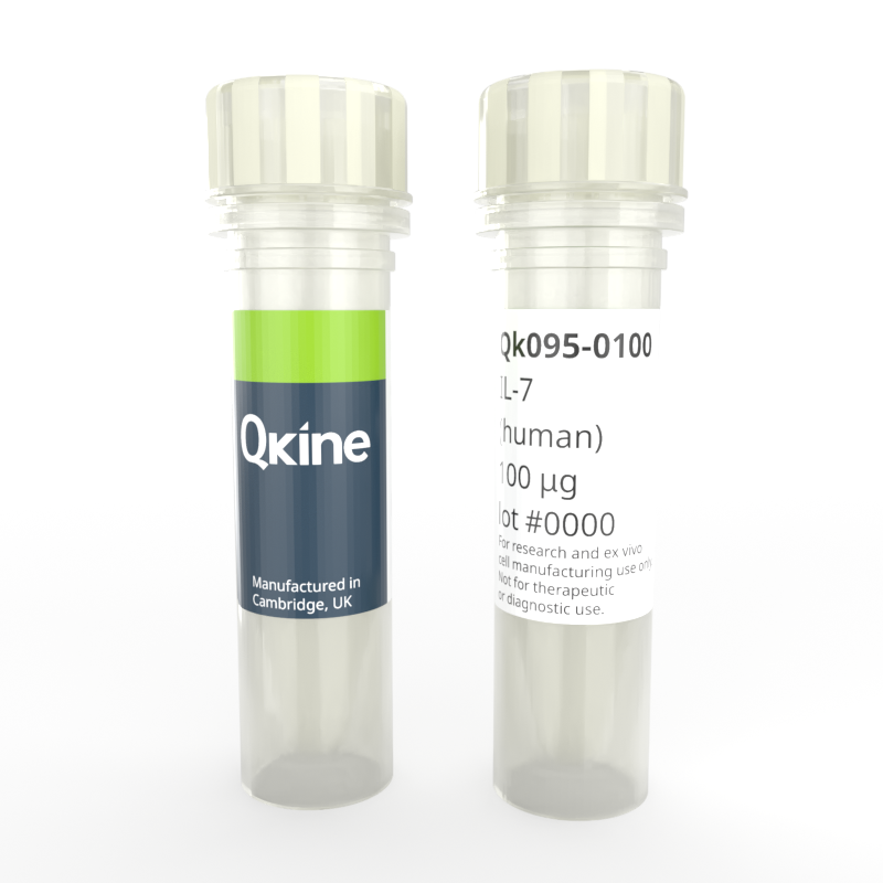
Recombinant human IL-7 protein
QK095
Brand: Qkine
Interleukin-7 (IL-7) is a vital cytokine essential for immune system regulation, particularly in the development and maintenance of T cells, playing a crucial role in both adaptive and innate immune responses. Recombinant human IL-7 stimulates the development of lymphoid progenitor cells.
Qkine has optimized the IL-7 manufacture process to produce a highly bioactive protein with excellent lot-to-lot consistency for enhanced experimental reproducibility. Qkine IL-7 is a highly pure 17.5 kDa monomer, animal origin-free (AOF) and carrier-protein-free (CF).

Currency:
| Product name | Catalog number | Pack size | Price | Price (USD) | Price (GBP) | Price (EUR) |
|---|---|---|---|---|---|---|
| Recombinant human IL-7 protein, 25 µg | QK095-0025 | 25 µg | (select above) | $ 340.00 | £ 250.00 | € 292.00 |
| Recombinant human IL-7 protein, 50 µg | QK095-0050 | 50 µg | (select above) | $ 520.00 | £ 380.00 | € 444.00 |
| Recombinant human IL-7 protein, 100 µg | QK095-0100 | 100 µg | (select above) | $ 685.00 | £ 500.00 | € 584.00 |
| Recombinant human IL-7 protein, 500 µg | QK095-0500 | 500 µg | (select above) | $ 2775.00 | £ 2025.00 | € 2366.00 |
| Recombinant human IL-7 , 1000 µg | QK095-1000 | 1000 µg | (select above) | $ 4370.00 | £ 3190.00 | € 3726.00 |
Note: prices shown do not include shipping and handling charges.
Qkine company name and logo are the property of Qkine Ltd. UK.
Alternative protein names
Species reactivity
human
species similarity:
mouse – 56%
rat – 57%
porcine – 69%
bovine – 71%
Summary
- High purity human IL-7 protein (Uniprot number: P13232)
- 17.5 kDa (monomer)
- >98%, by SDS-PAGE quantitative densitometry
- Expressed in E. coli
- Animal origin-free (AOF) and carrier protein-free
- Manufactured in Qkine's Cambridge, UK laboratories
- Lyophilized from acetonitrile, TFA
- Resuspend in sterile-filtered water at >50 µg/ml, add carrier protein if desired, prepare single use aliquots and store frozen at -20 °C (short-term) or -80 °C (long-term).
Featured applications
- Development, survival, and function of T cells
- Activation of T-cells and natural killer (NK) cells
- Development of regenerative medicine and vaccines
- Immunoregulatory function
Bioactivity
Recombinant IL-7 activity was determined using proliferation of mouse-derived B lymphocyte cell line 2E8. Cells were treated in duplicate with a serial dilution of IL-7 for 65 hours. Cell viability was measured using the CellTiter 96® Aqueous Non-Radioactive Cell Proliferation Assay (Promega). Data from Qk095 lot 204669. EC50 = 0.40 ng/ml (23 pM).

Purity
Recombinant IL-7 migrates as a major band at approximately 17.5 kDa (monomer) in reduced (R) and non-reduced (NR) conditions. No contaminating protein bands are present. The purified recombinant protein (3 µg) was resolved using 15% w/v SDS-PAGE in reduced (+β-mercaptoethanol, R) and non-reduced (NR) conditions and stained with Coomassie Brilliant Blue R250. Data from Qk095 lot #204669.

Further quality assays
- Mass spectrometry, single species with the expected mass
- Endotoxin: <0.005 EU/μg protein (below the level of detection)
- Recovery from stock vial: >95%

Qkine IL-7 is as biologically active as a comparable alternative supplier IL-7 protein. Recombinant IL-7 activity was determined using proliferation of the mouse-derived B lymphocyte cell line 2E8. Cells were treated in duplicate with a serial dilution of Qkine IL-7 (Qk095, green) for 65 hours. Cell viability was measured using the CellTiter 96 Aqueous Non-Radioactive Cell Proliferation Assay (Promega). Assay was performed by SBH Sciences using their standard (black) for comparison. Data from Qk095 lot #204669.
Protein background
IL-7 is a non-glycosylated protein with a molecular weight of about 17.5 kDa. It features four alpha-helices that facilitate its interaction with the IL-7 receptor (IL-7R), a heterodimer consisting of the IL-7Rα chain (CD127) and the common gamma chain (γc, CD132). This binding initiates signaling through the JAK-STAT pathway, particularly involving STAT5 phosphorylation, which promotes T cell survival, proliferation, and differentiation [1].
IL-7 is essential for the survival and homeostasis of T cells, supporting the development of thymocytes in the thymus and maintaining naive and memory T cells in the periphery, thus ensuring effective immune surveillance and long-term memory [2]. IL-7 influences B cell development and plays a role, though less prominently, in the development of natural killer (NK) cells [2].
IL-7 is extensively studied for its potential in cancer immunotherapy, where it enhances immune responses, particularly in promoting T cell recovery following chemotherapy or radiation, and in combination with checkpoint inhibitors [4]. In HIV and other chronic infections, IL-7 is explored for its ability to restore immune function by increasing CD4+ T cell counts and reducing immune exhaustion, potentially improving the effectiveness of existing therapies [3]. IL-7 also plays a key role in bone marrow transplantation, where it accelerates T cell recovery, reduces immune vulnerability, and is studied for its potential to mitigate graft-versus-host disease (GVHD) [4, 5].
In autoimmune diseases like multiple sclerosis and rheumatoid arthritis, IL-7 is a target for controlling autoreactive T cell survival and proliferation, offering new therapeutic approaches [5]. IL-7 is investigated as a vaccine adjuvant, particularly in vaccines requiring strong T cell responses, and in regenerative medicine, where its role in expanding T cells and hematopoietic stem cells is harnessed to enhance the efficacy of immune system regeneration therapies [4-6].
Background references
- McElroy, C.A., Holland, P.J., Zhao, P., et al. Structural reorganization of the interleukin-7 signaling complex. Proc. Natl Acad. Sci. USA 109, 2503–2508 (2012).
- Barata, J.T., Silva, A., Brandão, J.G., et al. IL-7 and IL-7R in immune homeostasis and cancer. Trends Immunol. 40, 580–594 (2019).
- Seddon, B., Tomlinson, P. & Zamoyska, R. Interleukin 7 and T cell receptor signals regulate homeostasis of CD4 memory cells. Nat. Immunol. 4, 680-686 (2003).
- Schluns, K.S., Kieper, W.C., Jameson, S.C. & Lefrançois, L. Interleukin-7 mediates the homeostasis of naïve and memory CD8 T cells in vivo. Nat. Immunol. 1, 426-432 (2000).
- Sportès, C. et al. Administration of rhIL-7 in humans increases in vivo TCR repertoire diversity by preferential expansion of naive T cell subsets. J. Exp. Med. 205, 1701-1714 (2008).
- Conlon, K.C. et al. IL-15 and IL-7 receptor alpha-dependent expansion of dual TCR T lymphocytes in patients with refractory malignancy receiving IL-15. J. Clin. Invest. 125, 94-110 (2015).
FAQ
What is IL-7?
IL-7 is an essential cytokine involved in the development, survival, and maintenance of T-cells, crucial for the immune response. It supports T-cell homeostasis, aids in B-cell development, and is being studied for therapeutic potential in immunodeficiency disorders, cancer treatment, and enhancing vaccine responses by boosting immune function.
Where is IL-7 found?
IL-7 is primarily produced by stromal cells in the bone marrow and thymus. It is also found in secondary lymphoid organs, such as lymph nodes, and is produced by non-lymphoid tissues like the skin, gut, and liver.
Is IL-7 a cytokine?
Yes, IL-7 is a cytokine. Cytokines are signaling molecules that mediate and regulate immunity, inflammation, and hematopoiesis.
What does the IL-7 gene do?
The IL-7 gene encodes the protein interleukin-7 (IL-7), which is a cytokine critical for the development and maintenance of the immune system, particularly in T cells.
What does IL-7 bind to?
IL-7 binds to the IL-7 receptor, composed of the IL-7Rα (CD127) and the common gamma chain (γc, CD132).
What is the function of the IL-7 receptor?
The IL-7 receptor (IL-7R) functions to mediate the survival, proliferation, and differentiation of T cells. It does this by binding IL-7 and triggering signaling pathways, particularly the JAK-STAT pathway, which promotes T cell development in the thymus, maintains peripheral T cell homeostasis, and supports early B cell development.
What is the IL-7 signaling pathway?
The IL-7 pathway involves IL-7 binding to its receptor, activating the JAK-STAT signaling cascade, which promotes T cell survival, proliferation, and differentiation.
How is IL-7 used in cell culture?
IL-7 is used to support the growth, survival, and proliferation of T cells and B cell precursors. It’s particularly vital in expanding T cells for research, immunotherapy, and studying immune cell development in vitro.